Media Center
Leading neuroscientist and AI geneticist Anders Dale, Ph.D. named president of J. Craig Venter Institute and joins Board
Through two multimillion-dollar NIH grants, Dr. Dale will continue leading key centers for the largest long-term studies of brain development and child health in the United States
Researchers design tools to develop vaccines more efficiently for African swine fever virus (ASFV)
The reverse-genetics system developed for ASFV may be adapted for other viruses, including lumpy skin disease, Zika, chikungunya, and Ebola viruses
Statement on cuts to National Institutes of Health funding
Disruptions or reductions in funding may irreparably harm biomedical research efforts at J. Craig Venter Institute and in the broader research ecosystem
Revolutionizing plastic waste management through biological upcycling
Innovative research transforms plastic waste into valuable chemicals, paving the way for a circular economy and sustainable space travel
Researchers call for global discussion about possible risks from “mirror bacteria”
Advocacy in Action: Effective Techniques for Shaping Science Policy
J. Craig Venter Institute awarded 5-year, $5M grant to lead Center for Innovative Recycling and Circular Economy (CIRCLE)
CIRCLE is one of the six new NSF Global Centers focused on advancing bioeconomy research to solve global challenges
Scientists discover molecular predictors of toxic algal blooms that pose health risk, ecological and economic harm
Genes in the algae Pseudo-nitzschia genus have been identified that act as a warning beacon for a dangerous neurotoxin
Prebys Introduces 2024 Grant Funding to Enhance Career Opportunities for Youth Across San Diego County
Organizations Receiving Part of the $5.89 Million Foster a Thriving San Diego Workforce
Prebys funds 24 grantees as part of a commitment to ensuring San Diego County youth are thriving and actively engaged in their communities
Passing of former J. Craig Venter Institute Trustee Bill Walton
Pages
Media Contact
Related
June Grant Update
Congratulations to our JCVI Principal Investigators for the several successful grants that were awarded or that we received notification of in the month of June. All of the following PIs received official confirmation of awards to be made to them. Christopher Dupont, John Glass, Granger...
Q&A with Jessie J. Knight, Jr.
The JCVI CEO Council is a small group of distinguished men and women who are thought leaders in business, medicine, law, the arts and humanities, and community affairs. JCVI is fortunate to have individuals willing to serve as knowledgeable and enthusiastic ambassadors for our scientists and...
JCVI Scientist Tackles Global Sanitation Challenges
Orianna Bretschger received her B.S. in Physics and Astronomy at the University of Northern Arizona. After a five- year career in aerospace and consulting, she completed a PhD in Materials Science at the University of Southern California. Eager to focus her efforts on alternative energy...
Dr. Venter Delivers UCSD 2015 School of Medicine Commencement
Full text for the address follows. J. Craig Venter, PhD, UCSD , 2015 School of Medicine Commencement Address Chancellor Khosla, Dean Brenner, Dean Savoia, UC Regent Charlene Zettel, UC Regent Sheldon Engelhorn, invited guests, families and graduates, thank you for inviting me...
Johns Hopkins Announces Inaugural Recipient of Hamilton Smith Award for Innovative Research
JCVI's Hamilton O. Smith, MD has been recognized by Johns Hopkins University with a research award in his honor. The inaugural recipient of the award is Jie Xiao, an associate professor of biophysics and biophysical chemistry at the Johns Hopkins University School of Medicine. Dr....
Meet Richard Scheuermann, Ph.D., JCVI’s Director of Bioinformatics
Richard H. Scheuermann, Ph.D., who joined JCVI in 2012 from the University of Texas Southwestern as the Director of Bioinformatics, is an accomplished researcher and educator. He and his team apply their deep knowledge in molecular immunology and infectious disease to develop novel...
Zoo in You Exhibit Now Open
Did you know trillions of microbes make their homes inside your body? In fact, these microorganisms outnumber our human cells 10 to 1, “colonize” us right from birth, and are so interwoven into our existence that without each other, none of us would survive! Thanks to new sophisticated...
In Memory of Dr. J. Robert Beyster
The JCVI family mourns the loss of a true friend and generous supporter, Dr. J. Robert Beyster. Dr. Beyster was a World War II Veteran, a nuclear engineer whose research propelled the Department of Defense's weapons systems and submarines into the future of war fighting, but most notably,...
Science on the Sea Ice Edge
On Sunday, December 14th JCVI scientists Andy Allen, Erin Bertrand, and Jeff Hoffman flew to New Zealand to begin the arduous journey to the sea ice edge of Antarctica. The JCVI team was joined by three members of the University of Southern California, led by David Hutchins, and three members...
Animal Forensics and Molecular Biology Techniques
A one-day high school workshop for New Hampton School’s Project Week Hosted by the J. Craig Venter Institute, Rockville, Maryland – March 11, 2015 Every March, the New Hampton School, an independent high school in New Hampshire, holds Project Week, an experiential...
Pages
Scientist renowned for study of adolescent brains named president of J. Craig Venter Institute
Anders Dale says he will move roughly $10 million in NIH funding from UCSD to JCVI.
Mirror Bacteria Research Poses Significant Risks, Dozens of Scientists Warn
Synthetic biologists make artificial cells, but one particular kind isn’t worth the risk.
Can CRISPR help stop African Swine Fever?
Gene editing could create a successful vaccine to protect against the viral disease that has killed close to 2 million pigs globally since 2021.
Getting Under the Skin
Amid an insulin crisis, one project aims to engineer microscopic insulin pumps out of a skin bacterium.
Planet Microbe
There are more organisms in the sea, a vital producer of oxygen on Earth, than planets and stars in the universe.
The Next Climate Change Calamity?: We’re Ruining the Microbiome, According to Human-Genome-Pioneer Craig Venter
In a new book (coauthored with Venter), a Vanity Fair contributor presents the oceanic evidence that human activity is altering the fabric of life on a microscopic scale.
Lessons from the Minimal Cell
“Despite reducing the sequence space of possible trajectories, we conclude that streamlining does not constrain fitness evolution and diversification of populations over time. Genome minimization may even create opportunities for evolutionary exploitation of essential genes, which are commonly observed to evolve more slowly.”
Even Synthetic Life Forms With a Tiny Genome Can Evolve
By watching “minimal” cells regain the fitness they lost, researchers are testing whether a genome can be too simple to evolve.
Privacy concerns sparked by human DNA accidentally collected in studies of other species
Two research teams warn that human genomic “bycatch” can reveal private information
Pages
Logos
The JCVI logo is presented in two formats: stacked and inline. Both are acceptable, with no preference towards either. Any use of the J. Craig Venter Institute logo or name must be cleared through the JCVI Marketing and Communications team. Please submit requests to info@jcvi.org.
To download, choose a version below, right-click, and select “save link as” or similar.
Images
Following are images of our facilities, research areas, and staff for use in news media, education, and noncommercial applications, given attribution noted with each image. If you require something that is not provided or would like to use the image in a commercial application please reach out to the JCVI Marketing and Communications team at info@jcvi.org.
Human Genome

The Diploid Genome Sequence of J. Craig Venter
gff2ps achieved another genome landmark to visualize the annotation of the first published human diploid genome, included as Poster S1 of “The Diploid Genome Sequence of J. Craig Venter” (Levy et al., PLoS Biology, 5(10):e254, 2007). Courtesy J.F. Abril / Computational Genomics Lab, Universitat de Barcelona (compgen.bio.ub.edu/Genome_Posters).

Annotation of the Celera Human Genome Assembly
We have drawn the map of the Human Genome with gff2ps. 22 autosomic, X and Y chromosomes were displayed in a big poster appearing as Figure 1 of “The Sequence of the Human Genome” (Venter et al., Science, 291(5507):1304-1351, 2001). The single chromosome pictures can be accessed from here to visualize the web version of the “Annotation of the Celera Human Genome Assembly” poster. Courtesy J.F. Abril / Computational Genomics Lab, Universitat de Barcelona (compgen.bio.ub.edu/Genome_Posters).
Synthetic Cell

J. Craig Venter, Ph.D. and Hamilton O. Smith, M.D.
Credit: J. Craig Venter Institute

Hamilton O. Smith, M.D. and Clyde A. Hutchison III, Ph.D.
Credit: J. Craig Venter Institute

J. Craig Venter, Ph.D.
Credit: Brett Shipe / J. Craig Venter Institute

Clyde A. Hutchison III, Ph.D.
Credit: J. Craig Venter Institute

John Glass, Ph.D.
Credit: J. Craig Venter Institute

Dan Gibson, Ph.D.
Credit: J. Craig Venter Institute

Carole Lartigue, Ph.D.
Credit: J. Craig Venter Institute

JCVI Synthetic Biology Team
Credit: J. Craig Venter Institute

Aggregated M. mycoides JCVI-syn1.0
Negatively stained transmission electron micrographs of aggregated M. mycoides JCVI-syn1.0. Cells using 1% uranyl acetate on pure carbon substrate visualized using JEOL 1200EX transmission electron microscope at 80 keV. Electron micrographs were provided by Tom Deerinck and Mark Ellisman of the National Center for Microscopy and Imaging Research at the University of California at San Diego.

Dividing M. mycoides JCVI-syn1.0
Negatively stained transmission electron micrographs of dividing M. mycoides JCVI-syn1.0. Freshly fixed cells were stained using 1% uranyl acetate on pure carbon substrate visualized using JEOL 1200EX transmission electron microscope at 80 keV. Electron micrographs were provided by Tom Deerinck and Mark Ellisman of the National Center for Microscopy and Imaging Research at the University of California at San Diego.

Scanning Electron Micrographs of M. mycoides JCVI-syn1
Scanning electron micrographs of M. mycoides JCVI-syn1. Samples were post-fixed in osmium tetroxide, dehydrated and critical point dried with CO2 , then visualized using a Hitachi SU6600 scanning electron microscope at 2.0 keV. Electron micrographs were provided by Tom Deerinck and Mark Ellisman of the National Center for Microscopy and Imaging Research at the University of California at San Diego.

Mycoplasma mycoides JCVI-syn1.0
Credit: J. Craig Venter Institute

The Assembly of a Synthetic M. mycoides Genome in Yeast
Credit: J. Craig Venter Institute

M. mycoides JCVI-syn 1.0 and WT M. mycoides
Credit: J. Craig Venter Institute

Creating Bacteria from Prokaryotic Genomes Engineered in Yeast
Credit: J. Craig Venter Institute
See more on the first self-replicating synthetic bacterial cell.
Minimal Cell

Minimal Cell — JCVI-syn3.0
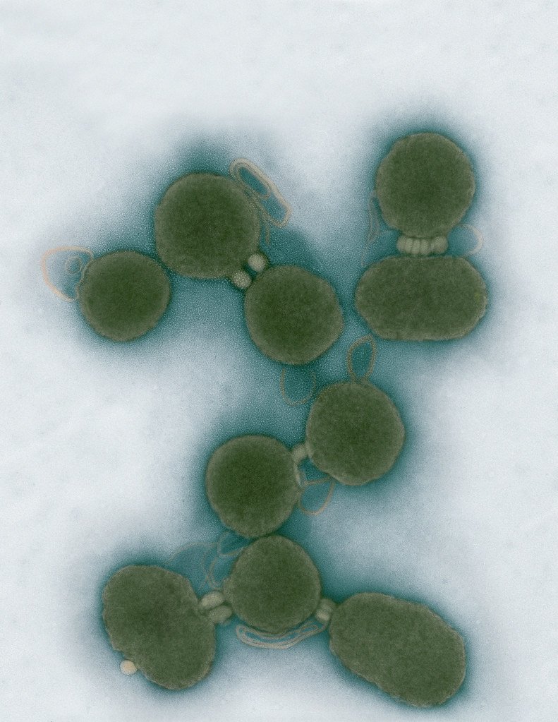
Electron micrographs of clusters of JCVI-syn3.0 cells magnified about 15,000 times. This is the world’s first minimal bacterial cell. Its synthetic genome contains only 473 genes. Surprisingly, the functions of 149 of those genes are unknown. The images were made by Tom Deerinck and Mark Ellisman of the National Center for Imaging and Microscopy Research at the University of California at San Diego.
Leadership

J. Craig Venter, Ph.D.
Credit: Brett Shipe / J. Craig Venter Institute

Sanjay Vashee, Ph.D.
Credit: J. Craig Venter Institute

John Glass, Ph.D.
Credit: J. Craig Venter Institute
Scientists in the Lab

JCVI Scientists Working in Lab
Credit: J. Craig Venter Institute

JCVI Scientists Working in Lab
Credit: J. Craig Venter Institute

JCVI Scientists Working in Lab
Credit: J. Craig Venter Institute

JCVI Scientists Working in Lab
Credit: J. Craig Venter Institute

JCVI Scientists Working in Lab
Credit: J. Craig Venter Institute

JCVI Scientists Working in Lab
Credit: J. Craig Venter Institute
JCVI La Jolla Lab (Exterior)

J. Craig Venter Institute, La Jolla (building exterior)
North facade at dusk. Nick Merrick © Hedrich Blessing Photographers.

J. Craig Venter Institute, La Jolla (building exterior)
South facade from soccer field. Nick Merrick © Hedrich Blessing Photographers.

J. Craig Venter Institute, La Jolla (building exterior)
Northwest view. Nick Merrick © Hedrich Blessing Photographers.

J. Craig Venter Institute, La Jolla (building exterior)
Northeast view of main entrance. Nick Merrick © Hedrich Blessing Photographers.

J. Craig Venter Institute, La Jolla (building exterior)
East facing main entrance at dusk. Nick Merrick © Hedrich Blessing Photographers.

J. Craig Venter Institute, La Jolla (building exterior)
East facing main entrance. Nick Merrick © Hedrich Blessing Photographers.

J. Craig Venter Institute, La Jolla (building exterior)
Building main entrance. Nick Merrick © Hedrich Blessing Photographers.

J. Craig Venter Institute, La Jolla (building exterior)
JCVI La Jolla north facade. Nick Merrick © Hedrich Blessing Photographers.

J. Craig Venter Institute, La Jolla (building exterior)
JCVI La Jolla north facade detail. Nick Merrick © Hedrich Blessing Photographers.

J. Craig Venter Institute, La Jolla (building exterior)
Rock garden in courtyard dusk. Nick Merrick © Hedrich Blessing Photographers.

J. Craig Venter Institute, La Jolla (building exterior)
Rock garden in courtyard. Nick Merrick © Hedrich Blessing Photographers.

J. Craig Venter Institute, La Jolla (building exterior)
Rock garden in courtyard. Nick Merrick © Hedrich Blessing Photographers.

J. Craig Venter Institute, La Jolla (building exterior)
People at courtyard tables. Nick Merrick © Hedrich Blessing Photographers.

J. Craig Venter Institute, La Jolla (building exterior)
2nd floor deck. © Tim Griffith.

J. Craig Venter Institute, La Jolla (building exterior)
Looking west at dusk. Nick Merrick © Hedrich Blessing Photographers.

J. Craig Venter Institute, La Jolla (building exterior)
First floor plaza looking south. Nick Merrick © Hedrich Blessing Photographers.

J. Craig Venter Institute, La Jolla (building exterior)
East main entrance closeup. Nick Merrick © Hedrich Blessing Photographers.

J. Craig Venter Institute, La Jolla (building exterior)
Stairs in courtyard. Nick Merrick © Hedrich Blessing Photographers.

J. Craig Venter Institute, La Jolla (building exterior)
Detail of southwest corner. Nick Merrick © Hedrich Blessing Photographers.

J. Craig Venter Institute, La Jolla (building exterior)
Sunset off 3rd floor deck. © Tim Griffith.

J. Craig Venter Institute, La Jolla (building exterior)
From northwest at dusk. Nick Merrick © Hedrich Blessing Photographers.

J. Craig Venter Institute, La Jolla (building exterior)
Photovoltaics looking west towards ocean. Nick Merrick © Hedrich Blessing Photographers.
JCVI La Jolla Lab (Interior)

J. Craig Venter Institute, La Jolla (building interior)
Wet lab with people. Nick Merrick © Hedrich Blessing Photographers.

J. Craig Venter Institute, La Jolla (building interior)
Single cell analyzer with researcher. © Tim Griffith.

J. Craig Venter Institute, La Jolla (building interior)
Mili-Q water purifier. © Tim Griffith.

J. Craig Venter Institute, La Jolla (building interior)
Lab bench work. Green plugs can be seen. © Tim Griffith.

J. Craig Venter Institute, La Jolla (building interior)
Cool room. © Tim Griffith.

J. Craig Venter Institute, La Jolla (building interior)
Confocal microscope. © Tim Griffith.

J. Craig Venter Institute, La Jolla (building interior)
Anaerobic glove box. © Tim Griffith.

J. Craig Venter Institute, La Jolla (building interior)
JCVI staff at DNA sequencer. © Tim Griffith.