Media Center
Schedule for 1998-1999 Distinguished Speaker Series announced.
TIGR announces the release of 1,937,631 bp of genome sequence from Chlorobium tepidum
Career Opportunities available at TIGR
The complete genome sequence of Treponema pallidum is published.
TIGR and the Naval Medical Research Institute announce the release of sequence from Plasmodium falciparum chromosome 2
TIGR announces the release of genome sequence from Porphyromonas gingivalis. license page.
Job Fair Announcement for the New Human Genome Venture
TIGR now offers technical training in genomic sequencing
TIGR announces the release of 3,209,119 bp of genome sequence from Enterococcus faecalis strain V583
Perkin-Elmer, Dr. Craig Venter, and TIGR Announce Formation of New Genomics Company
Plan to Sequence Human Genome Within Three Years
Pages
Media Contact
Related
Scientist Spotlight: Todd Michael
A love of science began for Todd Michael, PhD when his 7th grade teacher had him write a report on tree leaves. After collecting different leaves and looking up their tree type, he realized that although all of the trees were similar, they grew different types of leaves. He was certain there...
Fighting Back Against Flu
The 1918 influenza pandemic, which affected 500 million people globally and caused 50-100 million deaths, was the most severe pandemic in recorded history. Over the course of the last 100 years, advances in science and medicine have provided the tools to address influenza much more...
Scientist Spotlight: Marcelo Freire
Marcelo Freire, an associate professor in the Genomic Medicine and Infectious Disease Department at the J. Craig Venter Institute (JCVI), is currently working on decoding immune-microbiome genes and interactions. Growing up in Brazil and a curious person by nature, he often found himself...
Tracking Enterovirus D68, Cause of a Polio-like Illness in Some Patients
The J. Craig Venter Institute (JCVI) has played a vital role in defining the diversity of contemporary strains of human enteroviruses by using state-of-the art sequencing technologies, bioinformatics analyses, and in vitro and in vivo modeling.
Every Day is World Food Day at JCVI
World Food Day is a global initiative of the Food and Agriculture Organization (FAO) of the United Nations to ensure that people have access to enough high-quality food to lead active and healthy lives. After a period of decline, world hunger is on the rise again. Today, over 820 million people...
Mold Is Everywhere and Impacts You
When most people think about mold or fungi, food spoilage, a damp basement, or mushrooms come to mind. What you may not realize is how pervasive this branch of life is. Fungi is everywhere, from the ground you walk on to the air you breathe, and accounts for an estimated 25% of all biomass...
Scientists Discover Genetic Basis for Toxic Algal Blooms
Scientists from the J. Craig Venter Institute (JCVI) and Scripps Institution of Oceanography at the University of California San Diego have discovered how certain types of algal blooms become toxic, producing a harmful substance known as domoic acid. Microscopic view of domoic acid...
Ocean Microplastics Explained
As we wrap up sampling in the waters off of Maine, Dr. Chris Dupont discusses how collections of plastic particles in the water – or “plastisphere” – may be harboring fish or human pathogens. There may also be microbes responsible for degrading plastic, which are being investigated....
JCVI Team Awarded Two Grants Under the NSF’s “Understanding the Rules of Life” Initiative
The first award, led by John Glass, PhD, for $1M, is focused on “Building and Modeling Synthetic Bacterial Cells.” The second award, led by Zaida Luthey-Schulten, PhD, at the University of Illinois, also for $1M, is titled “Balancing the Demands of a Minimal Cell,” and is focused on...
Dr. Venter at Sailors’ Scuttlebutt Lecture Series
Dr. Craig Venter was a guest speaker at the Whaling Museum in partnership with Nantucket Community Sailing as part of the Sailors’ Scuttlebutt Lecture Series. Dr. Venter's lecture was titled, "Oceans, Human Health and the Genomic Future" discussing the Global Ocean...
Pages
Scientist renowned for study of adolescent brains named president of J. Craig Venter Institute
Anders Dale says he will move roughly $10 million in NIH funding from UCSD to JCVI.
Mirror Bacteria Research Poses Significant Risks, Dozens of Scientists Warn
Synthetic biologists make artificial cells, but one particular kind isn’t worth the risk.
Can CRISPR help stop African Swine Fever?
Gene editing could create a successful vaccine to protect against the viral disease that has killed close to 2 million pigs globally since 2021.
Getting Under the Skin
Amid an insulin crisis, one project aims to engineer microscopic insulin pumps out of a skin bacterium.
Planet Microbe
There are more organisms in the sea, a vital producer of oxygen on Earth, than planets and stars in the universe.
The Next Climate Change Calamity?: We’re Ruining the Microbiome, According to Human-Genome-Pioneer Craig Venter
In a new book (coauthored with Venter), a Vanity Fair contributor presents the oceanic evidence that human activity is altering the fabric of life on a microscopic scale.
Lessons from the Minimal Cell
“Despite reducing the sequence space of possible trajectories, we conclude that streamlining does not constrain fitness evolution and diversification of populations over time. Genome minimization may even create opportunities for evolutionary exploitation of essential genes, which are commonly observed to evolve more slowly.”
Even Synthetic Life Forms With a Tiny Genome Can Evolve
By watching “minimal” cells regain the fitness they lost, researchers are testing whether a genome can be too simple to evolve.
Privacy concerns sparked by human DNA accidentally collected in studies of other species
Two research teams warn that human genomic “bycatch” can reveal private information
Pages
Logos
The JCVI logo is presented in two formats: stacked and inline. Both are acceptable, with no preference towards either. Any use of the J. Craig Venter Institute logo or name must be cleared through the JCVI Marketing and Communications team. Please submit requests to info@jcvi.org.
To download, choose a version below, right-click, and select “save link as” or similar.
Images
Following are images of our facilities, research areas, and staff for use in news media, education, and noncommercial applications, given attribution noted with each image. If you require something that is not provided or would like to use the image in a commercial application please reach out to the JCVI Marketing and Communications team at info@jcvi.org.
Human Genome

The Diploid Genome Sequence of J. Craig Venter
gff2ps achieved another genome landmark to visualize the annotation of the first published human diploid genome, included as Poster S1 of “The Diploid Genome Sequence of J. Craig Venter” (Levy et al., PLoS Biology, 5(10):e254, 2007). Courtesy J.F. Abril / Computational Genomics Lab, Universitat de Barcelona (compgen.bio.ub.edu/Genome_Posters).

Annotation of the Celera Human Genome Assembly
We have drawn the map of the Human Genome with gff2ps. 22 autosomic, X and Y chromosomes were displayed in a big poster appearing as Figure 1 of “The Sequence of the Human Genome” (Venter et al., Science, 291(5507):1304-1351, 2001). The single chromosome pictures can be accessed from here to visualize the web version of the “Annotation of the Celera Human Genome Assembly” poster. Courtesy J.F. Abril / Computational Genomics Lab, Universitat de Barcelona (compgen.bio.ub.edu/Genome_Posters).
Synthetic Cell

J. Craig Venter, Ph.D. and Hamilton O. Smith, M.D.
Credit: J. Craig Venter Institute

Hamilton O. Smith, M.D. and Clyde A. Hutchison III, Ph.D.
Credit: J. Craig Venter Institute

J. Craig Venter, Ph.D.
Credit: Brett Shipe / J. Craig Venter Institute

Clyde A. Hutchison III, Ph.D.
Credit: J. Craig Venter Institute

John Glass, Ph.D.
Credit: J. Craig Venter Institute

Dan Gibson, Ph.D.
Credit: J. Craig Venter Institute

Carole Lartigue, Ph.D.
Credit: J. Craig Venter Institute

JCVI Synthetic Biology Team
Credit: J. Craig Venter Institute

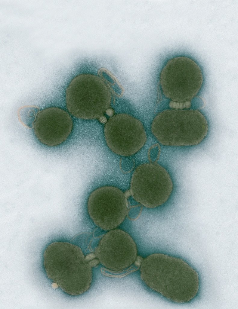
Aggregated M. mycoides JCVI-syn1.0
Negatively stained transmission electron micrographs of aggregated M. mycoides JCVI-syn1.0. Cells using 1% uranyl acetate on pure carbon substrate visualized using JEOL 1200EX transmission electron microscope at 80 keV. Electron micrographs were provided by Tom Deerinck and Mark Ellisman of the National Center for Microscopy and Imaging Research at the University of California at San Diego.

Dividing M. mycoides JCVI-syn1.0
Negatively stained transmission electron micrographs of dividing M. mycoides JCVI-syn1.0. Freshly fixed cells were stained using 1% uranyl acetate on pure carbon substrate visualized using JEOL 1200EX transmission electron microscope at 80 keV. Electron micrographs were provided by Tom Deerinck and Mark Ellisman of the National Center for Microscopy and Imaging Research at the University of California at San Diego.

Scanning Electron Micrographs of M. mycoides JCVI-syn1
Scanning electron micrographs of M. mycoides JCVI-syn1. Samples were post-fixed in osmium tetroxide, dehydrated and critical point dried with CO2 , then visualized using a Hitachi SU6600 scanning electron microscope at 2.0 keV. Electron micrographs were provided by Tom Deerinck and Mark Ellisman of the National Center for Microscopy and Imaging Research at the University of California at San Diego.

Mycoplasma mycoides JCVI-syn1.0
Credit: J. Craig Venter Institute

The Assembly of a Synthetic M. mycoides Genome in Yeast
Credit: J. Craig Venter Institute

M. mycoides JCVI-syn 1.0 and WT M. mycoides
Credit: J. Craig Venter Institute

Creating Bacteria from Prokaryotic Genomes Engineered in Yeast
Credit: J. Craig Venter Institute
See more on the first self-replicating synthetic bacterial cell.
Minimal Cell
Leadership

J. Craig Venter, Ph.D.
Credit: Brett Shipe / J. Craig Venter Institute

Sanjay Vashee, Ph.D.
Credit: J. Craig Venter Institute

John Glass, Ph.D.
Credit: J. Craig Venter Institute
Scientists in the Lab
JCVI La Jolla Lab (Exterior)

J. Craig Venter Institute, La Jolla (building exterior)
North facade at dusk. Nick Merrick © Hedrich Blessing Photographers.

J. Craig Venter Institute, La Jolla (building exterior)
South facade from soccer field. Nick Merrick © Hedrich Blessing Photographers.

J. Craig Venter Institute, La Jolla (building exterior)
Northwest view. Nick Merrick © Hedrich Blessing Photographers.

J. Craig Venter Institute, La Jolla (building exterior)
Northeast view of main entrance. Nick Merrick © Hedrich Blessing Photographers.

J. Craig Venter Institute, La Jolla (building exterior)
East facing main entrance at dusk. Nick Merrick © Hedrich Blessing Photographers.

J. Craig Venter Institute, La Jolla (building exterior)
East facing main entrance. Nick Merrick © Hedrich Blessing Photographers.

J. Craig Venter Institute, La Jolla (building exterior)
Building main entrance. Nick Merrick © Hedrich Blessing Photographers.

J. Craig Venter Institute, La Jolla (building exterior)
JCVI La Jolla north facade. Nick Merrick © Hedrich Blessing Photographers.

J. Craig Venter Institute, La Jolla (building exterior)
JCVI La Jolla north facade detail. Nick Merrick © Hedrich Blessing Photographers.

J. Craig Venter Institute, La Jolla (building exterior)
Rock garden in courtyard dusk. Nick Merrick © Hedrich Blessing Photographers.

J. Craig Venter Institute, La Jolla (building exterior)
Rock garden in courtyard. Nick Merrick © Hedrich Blessing Photographers.

J. Craig Venter Institute, La Jolla (building exterior)
Rock garden in courtyard. Nick Merrick © Hedrich Blessing Photographers.

J. Craig Venter Institute, La Jolla (building exterior)
People at courtyard tables. Nick Merrick © Hedrich Blessing Photographers.

J. Craig Venter Institute, La Jolla (building exterior)
2nd floor deck. © Tim Griffith.

J. Craig Venter Institute, La Jolla (building exterior)
Looking west at dusk. Nick Merrick © Hedrich Blessing Photographers.

J. Craig Venter Institute, La Jolla (building exterior)
First floor plaza looking south. Nick Merrick © Hedrich Blessing Photographers.

J. Craig Venter Institute, La Jolla (building exterior)
East main entrance closeup. Nick Merrick © Hedrich Blessing Photographers.

J. Craig Venter Institute, La Jolla (building exterior)
Stairs in courtyard. Nick Merrick © Hedrich Blessing Photographers.

J. Craig Venter Institute, La Jolla (building exterior)
Detail of southwest corner. Nick Merrick © Hedrich Blessing Photographers.

J. Craig Venter Institute, La Jolla (building exterior)
Sunset off 3rd floor deck. © Tim Griffith.

J. Craig Venter Institute, La Jolla (building exterior)
From northwest at dusk. Nick Merrick © Hedrich Blessing Photographers.

J. Craig Venter Institute, La Jolla (building exterior)
Photovoltaics looking west towards ocean. Nick Merrick © Hedrich Blessing Photographers.
JCVI La Jolla Lab (Interior)

J. Craig Venter Institute, La Jolla (building interior)
Wet lab with people. Nick Merrick © Hedrich Blessing Photographers.

J. Craig Venter Institute, La Jolla (building interior)
Single cell analyzer with researcher. © Tim Griffith.

J. Craig Venter Institute, La Jolla (building interior)
Mili-Q water purifier. © Tim Griffith.

J. Craig Venter Institute, La Jolla (building interior)
Lab bench work. Green plugs can be seen. © Tim Griffith.

J. Craig Venter Institute, La Jolla (building interior)
Cool room. © Tim Griffith.

J. Craig Venter Institute, La Jolla (building interior)
Confocal microscope. © Tim Griffith.

J. Craig Venter Institute, La Jolla (building interior)
Anaerobic glove box. © Tim Griffith.

J. Craig Venter Institute, La Jolla (building interior)
JCVI staff at DNA sequencer. © Tim Griffith.