Media Center
Logos
The JCVI logo is presented in two formats: stacked and inline. Both are acceptable, with no preference towards either. Any use of the J. Craig Venter Institute logo or name must be cleared through the JCVI Marketing and Communications team. Please submit requests to info@jcvi.org.
To download, choose a version below, right-click, and select “save link as” or similar.
Images
Following are images of our facilities, research areas, and staff for use in news media, education, and noncommercial applications, given attribution noted with each image. If you require something that is not provided or would like to use the image in a commercial application please reach out to the JCVI Marketing and Communications team at info@jcvi.org.
Human Genome

The Diploid Genome Sequence of J. Craig Venter
gff2ps achieved another genome landmark to visualize the annotation of the first published human diploid genome, included as Poster S1 of “The Diploid Genome Sequence of J. Craig Venter” (Levy et al., PLoS Biology, 5(10):e254, 2007). Courtesy J.F. Abril / Computational Genomics Lab, Universitat de Barcelona (compgen.bio.ub.edu/Genome_Posters).

Annotation of the Celera Human Genome Assembly
We have drawn the map of the Human Genome with gff2ps. 22 autosomic, X and Y chromosomes were displayed in a big poster appearing as Figure 1 of “The Sequence of the Human Genome” (Venter et al., Science, 291(5507):1304-1351, 2001). The single chromosome pictures can be accessed from here to visualize the web version of the “Annotation of the Celera Human Genome Assembly” poster. Courtesy J.F. Abril / Computational Genomics Lab, Universitat de Barcelona (compgen.bio.ub.edu/Genome_Posters).
Synthetic Cell

J. Craig Venter, Ph.D. and Hamilton O. Smith, M.D.
Credit: J. Craig Venter Institute

Hamilton O. Smith, M.D. and Clyde A. Hutchison III, Ph.D.
Credit: J. Craig Venter Institute

J. Craig Venter, Ph.D.
Credit: Brett Shipe / J. Craig Venter Institute

Clyde A. Hutchison III, Ph.D.
Credit: J. Craig Venter Institute

John Glass, Ph.D.
Credit: J. Craig Venter Institute

Dan Gibson, Ph.D.
Credit: J. Craig Venter Institute

Carole Lartigue, Ph.D.
Credit: J. Craig Venter Institute

JCVI Synthetic Biology Team
Credit: J. Craig Venter Institute
Aggregated M. mycoides JCVI-syn1.0
Negatively stained transmission electron micrographs of aggregated M. mycoides JCVI-syn1.0. Cells using 1% uranyl acetate on pure carbon substrate visualized using JEOL 1200EX transmission electron microscope at 80 keV. Electron micrographs were provided by Tom Deerinck and Mark Ellisman of the National Center for Microscopy and Imaging Research at the University of California at San Diego.

Dividing M. mycoides JCVI-syn1.0
Negatively stained transmission electron micrographs of dividing M. mycoides JCVI-syn1.0. Freshly fixed cells were stained using 1% uranyl acetate on pure carbon substrate visualized using JEOL 1200EX transmission electron microscope at 80 keV. Electron micrographs were provided by Tom Deerinck and Mark Ellisman of the National Center for Microscopy and Imaging Research at the University of California at San Diego.

Scanning Electron Micrographs of M. mycoides JCVI-syn1
Scanning electron micrographs of M. mycoides JCVI-syn1. Samples were post-fixed in osmium tetroxide, dehydrated and critical point dried with CO2 , then visualized using a Hitachi SU6600 scanning electron microscope at 2.0 keV. Electron micrographs were provided by Tom Deerinck and Mark Ellisman of the National Center for Microscopy and Imaging Research at the University of California at San Diego.

Mycoplasma mycoides JCVI-syn1.0
Credit: J. Craig Venter Institute

The Assembly of a Synthetic M. mycoides Genome in Yeast
Credit: J. Craig Venter Institute

M. mycoides JCVI-syn 1.0 and WT M. mycoides
Credit: J. Craig Venter Institute

Creating Bacteria from Prokaryotic Genomes Engineered in Yeast
Credit: J. Craig Venter Institute
See more on the first self-replicating synthetic bacterial cell.
Minimal Cell

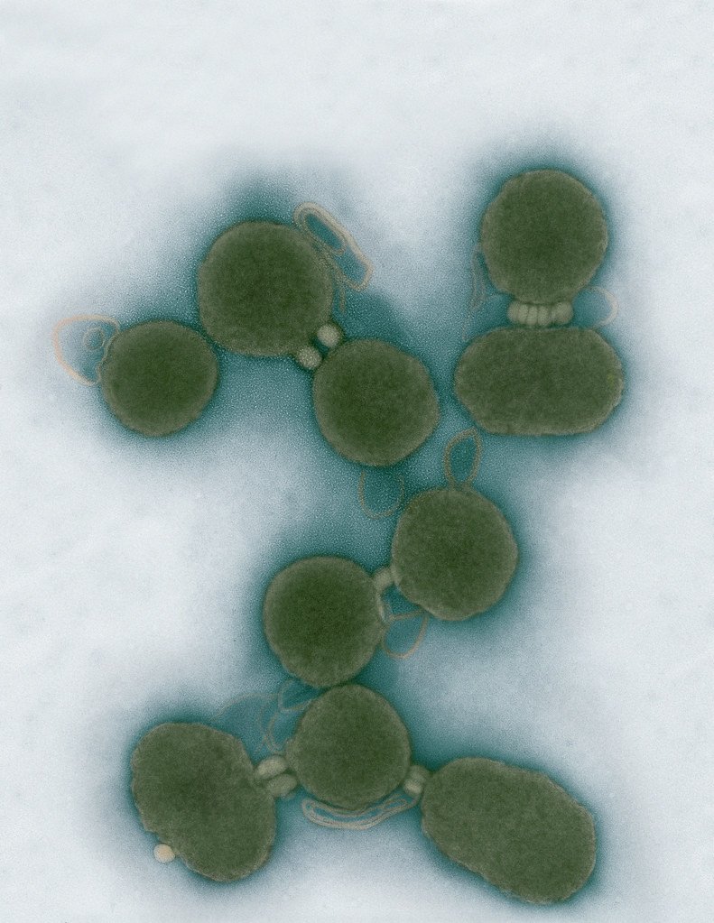
Minimal Cell — JCVI-syn3.0
Electron micrographs of clusters of JCVI-syn3.0 cells magnified about 15,000 times. This is the world’s first minimal bacterial cell. Its synthetic genome contains only 473 genes. Surprisingly, the functions of 149 of those genes are unknown. The images were made by Tom Deerinck and Mark Ellisman of the National Center for Imaging and Microscopy Research at the University of California at San Diego.
Leadership

J. Craig Venter, Ph.D.
Credit: Brett Shipe / J. Craig Venter Institute

Sanjay Vashee, Ph.D.
Credit: J. Craig Venter Institute

John Glass, Ph.D.
Credit: J. Craig Venter Institute
Scientists in the Lab

JCVI Scientists Working in Lab
Credit: J. Craig Venter Institute

JCVI Scientists Working in Lab
Credit: J. Craig Venter Institute

JCVI Scientists Working in Lab
Credit: J. Craig Venter Institute

JCVI Scientists Working in Lab
Credit: J. Craig Venter Institute

JCVI Scientists Working in Lab
Credit: J. Craig Venter Institute

JCVI Scientists Working in Lab
Credit: J. Craig Venter Institute
JCVI La Jolla Lab (Exterior)
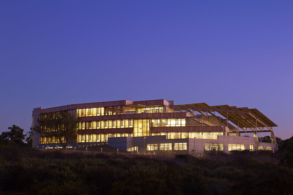
J. Craig Venter Institute, La Jolla (building exterior)
North facade at dusk. Nick Merrick © Hedrich Blessing Photographers.

J. Craig Venter Institute, La Jolla (building exterior)
South facade from soccer field. Nick Merrick © Hedrich Blessing Photographers.

J. Craig Venter Institute, La Jolla (building exterior)
Northwest view. Nick Merrick © Hedrich Blessing Photographers.

J. Craig Venter Institute, La Jolla (building exterior)
Northeast view of main entrance. Nick Merrick © Hedrich Blessing Photographers.

J. Craig Venter Institute, La Jolla (building exterior)
East facing main entrance at dusk. Nick Merrick © Hedrich Blessing Photographers.

J. Craig Venter Institute, La Jolla (building exterior)
East facing main entrance. Nick Merrick © Hedrich Blessing Photographers.

J. Craig Venter Institute, La Jolla (building exterior)
Building main entrance. Nick Merrick © Hedrich Blessing Photographers.

J. Craig Venter Institute, La Jolla (building exterior)
JCVI La Jolla north facade. Nick Merrick © Hedrich Blessing Photographers.

J. Craig Venter Institute, La Jolla (building exterior)
JCVI La Jolla north facade detail. Nick Merrick © Hedrich Blessing Photographers.

J. Craig Venter Institute, La Jolla (building exterior)
Rock garden in courtyard dusk. Nick Merrick © Hedrich Blessing Photographers.

J. Craig Venter Institute, La Jolla (building exterior)
Rock garden in courtyard. Nick Merrick © Hedrich Blessing Photographers.

J. Craig Venter Institute, La Jolla (building exterior)
Rock garden in courtyard. Nick Merrick © Hedrich Blessing Photographers.

J. Craig Venter Institute, La Jolla (building exterior)
People at courtyard tables. Nick Merrick © Hedrich Blessing Photographers.

J. Craig Venter Institute, La Jolla (building exterior)
2nd floor deck. © Tim Griffith.

J. Craig Venter Institute, La Jolla (building exterior)
Looking west at dusk. Nick Merrick © Hedrich Blessing Photographers.

J. Craig Venter Institute, La Jolla (building exterior)
First floor plaza looking south. Nick Merrick © Hedrich Blessing Photographers.

J. Craig Venter Institute, La Jolla (building exterior)
East main entrance closeup. Nick Merrick © Hedrich Blessing Photographers.

J. Craig Venter Institute, La Jolla (building exterior)
Stairs in courtyard. Nick Merrick © Hedrich Blessing Photographers.

J. Craig Venter Institute, La Jolla (building exterior)
Detail of southwest corner. Nick Merrick © Hedrich Blessing Photographers.

J. Craig Venter Institute, La Jolla (building exterior)
Sunset off 3rd floor deck. © Tim Griffith.

J. Craig Venter Institute, La Jolla (building exterior)
From northwest at dusk. Nick Merrick © Hedrich Blessing Photographers.

J. Craig Venter Institute, La Jolla (building exterior)
Photovoltaics looking west towards ocean. Nick Merrick © Hedrich Blessing Photographers.
JCVI La Jolla Lab (Interior)

J. Craig Venter Institute, La Jolla (building interior)
Wet lab with people. Nick Merrick © Hedrich Blessing Photographers.

J. Craig Venter Institute, La Jolla (building interior)
Single cell analyzer with researcher. © Tim Griffith.

J. Craig Venter Institute, La Jolla (building interior)
Mili-Q water purifier. © Tim Griffith.

J. Craig Venter Institute, La Jolla (building interior)
Lab bench work. Green plugs can be seen. © Tim Griffith.

J. Craig Venter Institute, La Jolla (building interior)
Cool room. © Tim Griffith.

J. Craig Venter Institute, La Jolla (building interior)
Confocal microscope. © Tim Griffith.

J. Craig Venter Institute, La Jolla (building interior)
Anaerobic glove box. © Tim Griffith.

J. Craig Venter Institute, La Jolla (building interior)
JCVI staff at DNA sequencer. © Tim Griffith.