Media Center
J. Craig Venter Institute scientists awarded five-year, $5.7M grant from NIH to develop phage treatment
Phage research accelerates with the rise of antibiotic resistance to address increasingly prevalent and difficult to treat bacterial infections
Bringing cells to life … and to Minecraft: $30 million NSF grant to support whole-cell modeling at the Beckman Institute
Beckman researchers and collaborators received $30 million from the U.S. National Science Foundation to establish the NSF Science and Technology Center for Quantitative Cell Biology. The center will develop whole-cell models to transform our understanding of how cells function and share that knowledge with diverse communities through the popular computer game Minecraft.
NLM Selects Dr. Richard H. Scheuermann as Scientific Director for the National Library of Medicine
PolyBio Research Foundation Consortium & colleagues publishes Nature position paper on SARS-CoV-2 reservoir in Long COVID/PASC
Using AI to speed up vaccine development against Disease X
Artificial cells demonstrate that “life finds a way”
Family resemblance: How T cells could fight many coronaviruses at once
LJI researchers work to head off future pandemics by uncovering key similarities between SARS-CoV-2 and common cold coronaviruses
Scientists aim to develop vaccine against all deadly coronaviruses
$8 million NIH grant supports effort to avert next pandemic
What are the Drivers of Chronic Infectious Disease?
A $1 million grant from the Steven & Alexandra Cohen Foundation will launch a UC San Diego-led national effort to more deeply study tissue samples from patients with conditions ranging from long COVID-19 and relapsed Lyme disease to chronic fatigue syndrome
The Tissue Analysis Pipeline will be directed by scientists at UC San Diego and the J. Craig Venter Institute
BullFrog AI Partners with J. Craig Venter Institute to Develop Colorectal Cancer Therapeutic
Collaboration seeks to develop an oncolytic virus that incorporates a novel, precision-targeted approach to improve safety and efficacy
Pages
Media Contact
Related
Venter Institute Researchers Tackle the Growing Concern of Antibiotic Resistant Bacterial Infections with Genomic, Phage Approaches
The Centers for Disease Control and Prevention (CDC) estimates that each year in the United States two million people acquire antibiotic resistant bacterial infections that lead to 23,000 deaths. Antibiotic resistance affects people of all ages and seriously impacts the healthcare, veterinary,...
2019 Summer Internship Program
The 2019 Summer Internship Program which wrapped up in August was another rousing success at the J. Craig Venter Institute. Faculty and staff in both the Rockville (MD) and La Jolla (CA) campuses mentored and trained 25 students (high school, undergraduate, and graduate students)...
Diatoms Have Found a Way to Pirate Bacterial Iron Sources
In large regions of the world’s oceans, photosynthesis struggles to operate because a key ingredient is missing. Many of the proteins involved in harvesting energy from sunlight require iron atoms to function, but iron is hard to find in seawater. Most of the ocean is far removed from...
The JCVI Genomic Frontier Fund
As we complete our 26th year as a private genomic research institution, we are still just as excited as we were in the very beginning to be making new discoveries, potentially ones that will change our society for the better. The knowledge gained from our study of DNA, or as Dr. Venter...
New Sequencing Technologies Enable Better and Faster Understanding of the Human Microbiome
Humans have trillions of different species of microorganisms living inside and on the human body. These microbes colonize on the skin, gut, oral cavity, vagina, internal organs, and circulating fluids, and are called the human microbiome. The human microbiome plays profound roles in health...
Human Microbiome Research has Massive Potential for Health Applications
Thirteen years ago, a team led by J. Craig Venter Institute President, Karen Nelson, Ph.D., published the first major human microbiome study, radically changing the way we look at human health and the role the microbes that inhabit each of us play in disease. This seminal publication...
Scientist Spotlight: Lauren Oldfield
Since high school, Lauren Oldfield, PhD found that science was her calling. It started with a love of reading encouraged by her mom and grandmother, both avid readers, and weekly trips to the public library. Books by Michael Crichton and Richard Preston were staples in her grandmother’s...
When Starved, Dangerous Oral Bacteria Hang On
J. Craig Venter Institute (JCVI) postdoctoral fellow, Jonathon Baker, PhD and a team of researchers from JCVI, University of Washington, the University of California, Los Angeles, and The Forsyth Institute recently published their findings from the first study to examine the ecological dynamics...
No More Needles! Using Microbiome and Synthetic Biology Advances to Better Treat Type 1 Diabetes
Learn about exciting advances made by JCVI researchers Yo Suzuki and John Glass who are on a quest to better understand and treat Type 1 Diabetes (T1D). Currently T1D is managed by injecting insulin to manage blood glucose levels. Drs. Suzuki and Glass want to change that by creating a...
How to Bake a (Fungal) Turkey
From the kitchen of Stephanie Mounaud, Scientific Project Manager at JCVI Ingredients Media base (see media recipe) Agar Aspergillus terreus (multiple strains) Aspergillus niger Aspergillus fumigatus Aspergillus oryzae...
Pages
Public Health is the Next Big Thing at UC San Diego
Researchers have swapped the genome of gut germ E. coli for an artificial one
By creating a new genome, scientists could create organisms tailored to produce desirable compounds
Genetically modified bacteria-killing viruses used on patient for first time
Hair claimed to belong to Leonardo da Vinci to undergo DNA testing
Critics, however, argue that this effort is flawed from the beginning
Students learn about genomics, a life in science, at J. Craig Venter Institute
Pages
Logos
The JCVI logo is presented in two formats: stacked and inline. Both are acceptable, with no preference towards either. Any use of the J. Craig Venter Institute logo or name must be cleared through the JCVI Marketing and Communications team. Please submit requests to info@jcvi.org.
To download, choose a version below, right-click, and select “save link as” or similar.
Images
Following are images of our facilities, research areas, and staff for use in news media, education, and noncommercial applications, given attribution noted with each image. If you require something that is not provided or would like to use the image in a commercial application please reach out to the JCVI Marketing and Communications team at info@jcvi.org.
Human Genome

The Diploid Genome Sequence of J. Craig Venter
gff2ps achieved another genome landmark to visualize the annotation of the first published human diploid genome, included as Poster S1 of “The Diploid Genome Sequence of J. Craig Venter” (Levy et al., PLoS Biology, 5(10):e254, 2007). Courtesy J.F. Abril / Computational Genomics Lab, Universitat de Barcelona (compgen.bio.ub.edu/Genome_Posters).

Annotation of the Celera Human Genome Assembly
We have drawn the map of the Human Genome with gff2ps. 22 autosomic, X and Y chromosomes were displayed in a big poster appearing as Figure 1 of “The Sequence of the Human Genome” (Venter et al., Science, 291(5507):1304-1351, 2001). The single chromosome pictures can be accessed from here to visualize the web version of the “Annotation of the Celera Human Genome Assembly” poster. Courtesy J.F. Abril / Computational Genomics Lab, Universitat de Barcelona (compgen.bio.ub.edu/Genome_Posters).
Synthetic Cell

J. Craig Venter, Ph.D. and Hamilton O. Smith, M.D.
Credit: J. Craig Venter Institute

Hamilton O. Smith, M.D. and Clyde A. Hutchison III, Ph.D.
Credit: J. Craig Venter Institute

J. Craig Venter, Ph.D.
Credit: Brett Shipe / J. Craig Venter Institute

Clyde A. Hutchison III, Ph.D.
Credit: J. Craig Venter Institute

John Glass, Ph.D.
Credit: J. Craig Venter Institute

Dan Gibson, Ph.D.
Credit: J. Craig Venter Institute

Carole Lartigue, Ph.D.
Credit: J. Craig Venter Institute

JCVI Synthetic Biology Team
Credit: J. Craig Venter Institute

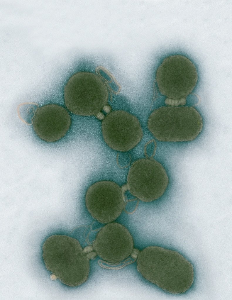
Aggregated M. mycoides JCVI-syn1.0
Negatively stained transmission electron micrographs of aggregated M. mycoides JCVI-syn1.0. Cells using 1% uranyl acetate on pure carbon substrate visualized using JEOL 1200EX transmission electron microscope at 80 keV. Electron micrographs were provided by Tom Deerinck and Mark Ellisman of the National Center for Microscopy and Imaging Research at the University of California at San Diego.

Dividing M. mycoides JCVI-syn1.0
Negatively stained transmission electron micrographs of dividing M. mycoides JCVI-syn1.0. Freshly fixed cells were stained using 1% uranyl acetate on pure carbon substrate visualized using JEOL 1200EX transmission electron microscope at 80 keV. Electron micrographs were provided by Tom Deerinck and Mark Ellisman of the National Center for Microscopy and Imaging Research at the University of California at San Diego.

Scanning Electron Micrographs of M. mycoides JCVI-syn1
Scanning electron micrographs of M. mycoides JCVI-syn1. Samples were post-fixed in osmium tetroxide, dehydrated and critical point dried with CO2 , then visualized using a Hitachi SU6600 scanning electron microscope at 2.0 keV. Electron micrographs were provided by Tom Deerinck and Mark Ellisman of the National Center for Microscopy and Imaging Research at the University of California at San Diego.

Mycoplasma mycoides JCVI-syn1.0
Credit: J. Craig Venter Institute

The Assembly of a Synthetic M. mycoides Genome in Yeast
Credit: J. Craig Venter Institute

M. mycoides JCVI-syn 1.0 and WT M. mycoides
Credit: J. Craig Venter Institute

Creating Bacteria from Prokaryotic Genomes Engineered in Yeast
Credit: J. Craig Venter Institute
See more on the first self-replicating synthetic bacterial cell.
Minimal Cell

Minimal Cell — JCVI-syn3.0
Electron micrographs of clusters of JCVI-syn3.0 cells magnified about 15,000 times. This is the world’s first minimal bacterial cell. Its synthetic genome contains only 473 genes. Surprisingly, the functions of 149 of those genes are unknown. The images were made by Tom Deerinck and Mark Ellisman of the National Center for Imaging and Microscopy Research at the University of California at San Diego.
Leadership

J. Craig Venter, Ph.D.
Credit: Brett Shipe / J. Craig Venter Institute

Sanjay Vashee, Ph.D.
Credit: J. Craig Venter Institute

John Glass, Ph.D.
Credit: J. Craig Venter Institute
Scientists in the Lab

JCVI Scientists Working in Lab
Credit: J. Craig Venter Institute

JCVI Scientists Working in Lab
Credit: J. Craig Venter Institute

JCVI Scientists Working in Lab
Credit: J. Craig Venter Institute

JCVI Scientists Working in Lab
Credit: J. Craig Venter Institute

JCVI Scientists Working in Lab
Credit: J. Craig Venter Institute

JCVI Scientists Working in Lab
Credit: J. Craig Venter Institute
JCVI La Jolla Lab (Exterior)

J. Craig Venter Institute, La Jolla (building exterior)
North facade at dusk. Nick Merrick © Hedrich Blessing Photographers.

J. Craig Venter Institute, La Jolla (building exterior)
South facade from soccer field. Nick Merrick © Hedrich Blessing Photographers.

J. Craig Venter Institute, La Jolla (building exterior)
Northwest view. Nick Merrick © Hedrich Blessing Photographers.

J. Craig Venter Institute, La Jolla (building exterior)
Northeast view of main entrance. Nick Merrick © Hedrich Blessing Photographers.

J. Craig Venter Institute, La Jolla (building exterior)
East facing main entrance at dusk. Nick Merrick © Hedrich Blessing Photographers.

J. Craig Venter Institute, La Jolla (building exterior)
East facing main entrance. Nick Merrick © Hedrich Blessing Photographers.

J. Craig Venter Institute, La Jolla (building exterior)
Building main entrance. Nick Merrick © Hedrich Blessing Photographers.

J. Craig Venter Institute, La Jolla (building exterior)
JCVI La Jolla north facade. Nick Merrick © Hedrich Blessing Photographers.

J. Craig Venter Institute, La Jolla (building exterior)
JCVI La Jolla north facade detail. Nick Merrick © Hedrich Blessing Photographers.

J. Craig Venter Institute, La Jolla (building exterior)
Rock garden in courtyard dusk. Nick Merrick © Hedrich Blessing Photographers.

J. Craig Venter Institute, La Jolla (building exterior)
Rock garden in courtyard. Nick Merrick © Hedrich Blessing Photographers.

J. Craig Venter Institute, La Jolla (building exterior)
Rock garden in courtyard. Nick Merrick © Hedrich Blessing Photographers.

J. Craig Venter Institute, La Jolla (building exterior)
People at courtyard tables. Nick Merrick © Hedrich Blessing Photographers.

J. Craig Venter Institute, La Jolla (building exterior)
2nd floor deck. © Tim Griffith.

J. Craig Venter Institute, La Jolla (building exterior)
Looking west at dusk. Nick Merrick © Hedrich Blessing Photographers.

J. Craig Venter Institute, La Jolla (building exterior)
First floor plaza looking south. Nick Merrick © Hedrich Blessing Photographers.

J. Craig Venter Institute, La Jolla (building exterior)
East main entrance closeup. Nick Merrick © Hedrich Blessing Photographers.

J. Craig Venter Institute, La Jolla (building exterior)
Stairs in courtyard. Nick Merrick © Hedrich Blessing Photographers.

J. Craig Venter Institute, La Jolla (building exterior)
Detail of southwest corner. Nick Merrick © Hedrich Blessing Photographers.

J. Craig Venter Institute, La Jolla (building exterior)
Sunset off 3rd floor deck. © Tim Griffith.

J. Craig Venter Institute, La Jolla (building exterior)
From northwest at dusk. Nick Merrick © Hedrich Blessing Photographers.

J. Craig Venter Institute, La Jolla (building exterior)
Photovoltaics looking west towards ocean. Nick Merrick © Hedrich Blessing Photographers.
JCVI La Jolla Lab (Interior)

J. Craig Venter Institute, La Jolla (building interior)
Wet lab with people. Nick Merrick © Hedrich Blessing Photographers.

J. Craig Venter Institute, La Jolla (building interior)
Single cell analyzer with researcher. © Tim Griffith.

J. Craig Venter Institute, La Jolla (building interior)
Mili-Q water purifier. © Tim Griffith.

J. Craig Venter Institute, La Jolla (building interior)
Lab bench work. Green plugs can be seen. © Tim Griffith.

J. Craig Venter Institute, La Jolla (building interior)
Cool room. © Tim Griffith.

J. Craig Venter Institute, La Jolla (building interior)
Confocal microscope. © Tim Griffith.

J. Craig Venter Institute, La Jolla (building interior)
Anaerobic glove box. © Tim Griffith.

J. Craig Venter Institute, La Jolla (building interior)
JCVI staff at DNA sequencer. © Tim Griffith.