Media Center
JCVI Associate Professor Marcelo Freire elected to the 2022 class of AAAS Fellows
More Than 500 Scientists and Engineers Bestowed Lifetime AAAS Fellows Honor
Therapeutic Potential of Bizarre ‘Jumbo’ Viruses Tapped for $10M HHMI Emerging Pathogens Project
UC San Diego leads initiative aiming to develop bacteriophages as solutions for antibiotic resistance crisis
HHMI’s Emerging Pathogens Initiative Aims for Scientific Head Start on Future Epidemics
Synthetic genomics advances and promise
Advances in DNA synthesis will enable extraordinary new opportunities in medicine, industry, agriculture, and research
Scientists announce comprehensive regional diagnostic of microbial ocean life using DNA testing
Large-scale ‘metabarcoding’ methods could revolutionize how society understands forces that drive seafood supply, planet’s ability to remove greenhouse gases
J. Craig Venter Institute sells La Jolla laboratory building to UC San Diego
2022 Fellows of the AACR Academy will be honored during Sunday’s Opening Session
JCVI Professor Emeritus Hamilton O. Smith, MD among the inductees
Scientists develop most complete whole-cell computer simulation model of cell to date
J. Craig Venter Institute model organism-minimal cell platform provides robust tools for exploring first principles of life, design tools for genome
Omicron and Beta variants evade antibodies elicited by vaccines and previous infections, but boosters help
Pregnancy also contributes to a reduced COVID-19 antibody response
Pages
Media Contact
Related
Heading north with more daylight
After spending a couple of days visiting with my family in Stockholm, I boarded a ferry boat to Blidö and rejoined the Sorcerer II crew to head north to the Bothnian Sea. Before departing, we sampled in the bay outside Dr. Norrby’s summer house. The last days of fantastic summer weather had...
The last leg of the Volvo Ocean Race, the Swedish Archipelago and the Gulf of Bothnia Sampling Transect
The morning of June 25th we left Stockholm and followed the Volvo race boats into the Baltic to watch the start of the last leg of the race to St. Petersburg. Once again there were hundreds of boats on the water to watch the start of the race. As the race began we saw someone waving to Dr....
In the News
We docked in the Volvo Ocean Race Village for a week. It was very exciting to be so close to all of the activities surrounding the race. Over the week Dr. Venter and Karolina and I were interviewed by many local and national TV, radio stations and newspapers. Here are some links to a few of the...
The Volvo Ocean Race
We arrived in Sandhamn at 10 p.m. on June 15th. It was perfect timing because the Volvo Ocean Race boats were arriving around 11 p.m. The Volvo Ocean Race, formally known as the Whitbread “Around the World Race,” began in Alicante on October 11th 2008 and ends in St. Petersburg on June 25th...
Heading to the Mother Land — Sweden
After transiting through the Kiel Canal, the waterway that links the North Sea to the Baltic Sea, and welcoming Dr. Venter in a rainy Copenhagen, we embarked for Sweden, my home and one of the main destinations of our 2009 expedition. It was a proud and special moment for me when first mate,...
Sampling in Helgoland — A warm German welcome for the Sorcerer II
After a little more than two weeks in Plymouth, UK the Sorcerer II set sail on June 3rd. We were sad to say goodbye to our new friends at PLM, but we were grateful for their hospitality, friendship and scientific collaboration. We're looking forward to coming back through Plymouth in the fall....
Cornish Pasties and Jellyfish at the MBA
On Monday we were invited to the Marine Biology Association (MBA) and the Sir Alister Hardy Foundation for Ocean Science (SAHFOS) for lunch and a more extensive tour of the laboratories and SAHFOS. This was an excellent opportunity for crew members who missed the first tour. A beautiful table...
The Final Plymouth Sample
On Thursday, May 28th the Sorcerer II crew, accompanied by Dr. Jack Gilbert and two of his PhD students, headed out for one final sampling trip. The destination was E-1, a long term research station for PML located about 25 miles off the coast of Plymouth in the English Channel. As we...
First Sampling in Plymouth Reveals Interesting Blooms — BBC Cameras capture it all!
After a couple of days in Plymouth we were ready for the first of two intense sampling days together with the Plymouth Marine Laboratory (PML). We had heard rumours about blooms of Phaeocystis, a conspicuous bloom-former in the North Sea and English Channel. When it blooms, it turns the...
Days of Discovery: Plymouth, Sea Urchin Cell Division and More Plankton
After a few days of fairly rough weather and winds up to 50 knots we finally spotted land and made our way to Plymouth. With our social interactions having been restricted to a pod of pilot whales and a few tankers passing through the night, we were excited to see a welcoming committee,...
Pages
Scientist renowned for study of adolescent brains named president of J. Craig Venter Institute
Anders Dale says he will move roughly $10 million in NIH funding from UCSD to JCVI.
Mirror Bacteria Research Poses Significant Risks, Dozens of Scientists Warn
Synthetic biologists make artificial cells, but one particular kind isn’t worth the risk.
Can CRISPR help stop African Swine Fever?
Gene editing could create a successful vaccine to protect against the viral disease that has killed close to 2 million pigs globally since 2021.
Getting Under the Skin
Amid an insulin crisis, one project aims to engineer microscopic insulin pumps out of a skin bacterium.
Planet Microbe
There are more organisms in the sea, a vital producer of oxygen on Earth, than planets and stars in the universe.
The Next Climate Change Calamity?: We’re Ruining the Microbiome, According to Human-Genome-Pioneer Craig Venter
In a new book (coauthored with Venter), a Vanity Fair contributor presents the oceanic evidence that human activity is altering the fabric of life on a microscopic scale.
Lessons from the Minimal Cell
“Despite reducing the sequence space of possible trajectories, we conclude that streamlining does not constrain fitness evolution and diversification of populations over time. Genome minimization may even create opportunities for evolutionary exploitation of essential genes, which are commonly observed to evolve more slowly.”
Even Synthetic Life Forms With a Tiny Genome Can Evolve
By watching “minimal” cells regain the fitness they lost, researchers are testing whether a genome can be too simple to evolve.
Privacy concerns sparked by human DNA accidentally collected in studies of other species
Two research teams warn that human genomic “bycatch” can reveal private information
Pages
Logos
The JCVI logo is presented in two formats: stacked and inline. Both are acceptable, with no preference towards either. Any use of the J. Craig Venter Institute logo or name must be cleared through the JCVI Marketing and Communications team. Please submit requests to info@jcvi.org.
To download, choose a version below, right-click, and select “save link as” or similar.
Images
Following are images of our facilities, research areas, and staff for use in news media, education, and noncommercial applications, given attribution noted with each image. If you require something that is not provided or would like to use the image in a commercial application please reach out to the JCVI Marketing and Communications team at info@jcvi.org.
Human Genome

The Diploid Genome Sequence of J. Craig Venter
gff2ps achieved another genome landmark to visualize the annotation of the first published human diploid genome, included as Poster S1 of “The Diploid Genome Sequence of J. Craig Venter” (Levy et al., PLoS Biology, 5(10):e254, 2007). Courtesy J.F. Abril / Computational Genomics Lab, Universitat de Barcelona (compgen.bio.ub.edu/Genome_Posters).

Annotation of the Celera Human Genome Assembly
We have drawn the map of the Human Genome with gff2ps. 22 autosomic, X and Y chromosomes were displayed in a big poster appearing as Figure 1 of “The Sequence of the Human Genome” (Venter et al., Science, 291(5507):1304-1351, 2001). The single chromosome pictures can be accessed from here to visualize the web version of the “Annotation of the Celera Human Genome Assembly” poster. Courtesy J.F. Abril / Computational Genomics Lab, Universitat de Barcelona (compgen.bio.ub.edu/Genome_Posters).
Synthetic Cell

J. Craig Venter, Ph.D. and Hamilton O. Smith, M.D.
Credit: J. Craig Venter Institute

Hamilton O. Smith, M.D. and Clyde A. Hutchison III, Ph.D.
Credit: J. Craig Venter Institute

J. Craig Venter, Ph.D.
Credit: Brett Shipe / J. Craig Venter Institute

Clyde A. Hutchison III, Ph.D.
Credit: J. Craig Venter Institute

John Glass, Ph.D.
Credit: J. Craig Venter Institute

Dan Gibson, Ph.D.
Credit: J. Craig Venter Institute

Carole Lartigue, Ph.D.
Credit: J. Craig Venter Institute

JCVI Synthetic Biology Team
Credit: J. Craig Venter Institute

Aggregated M. mycoides JCVI-syn1.0
Negatively stained transmission electron micrographs of aggregated M. mycoides JCVI-syn1.0. Cells using 1% uranyl acetate on pure carbon substrate visualized using JEOL 1200EX transmission electron microscope at 80 keV. Electron micrographs were provided by Tom Deerinck and Mark Ellisman of the National Center for Microscopy and Imaging Research at the University of California at San Diego.

Dividing M. mycoides JCVI-syn1.0
Negatively stained transmission electron micrographs of dividing M. mycoides JCVI-syn1.0. Freshly fixed cells were stained using 1% uranyl acetate on pure carbon substrate visualized using JEOL 1200EX transmission electron microscope at 80 keV. Electron micrographs were provided by Tom Deerinck and Mark Ellisman of the National Center for Microscopy and Imaging Research at the University of California at San Diego.

Scanning Electron Micrographs of M. mycoides JCVI-syn1
Scanning electron micrographs of M. mycoides JCVI-syn1. Samples were post-fixed in osmium tetroxide, dehydrated and critical point dried with CO2 , then visualized using a Hitachi SU6600 scanning electron microscope at 2.0 keV. Electron micrographs were provided by Tom Deerinck and Mark Ellisman of the National Center for Microscopy and Imaging Research at the University of California at San Diego.

Mycoplasma mycoides JCVI-syn1.0
Credit: J. Craig Venter Institute

The Assembly of a Synthetic M. mycoides Genome in Yeast
Credit: J. Craig Venter Institute

M. mycoides JCVI-syn 1.0 and WT M. mycoides
Credit: J. Craig Venter Institute

Creating Bacteria from Prokaryotic Genomes Engineered in Yeast
Credit: J. Craig Venter Institute
See more on the first self-replicating synthetic bacterial cell.
Minimal Cell

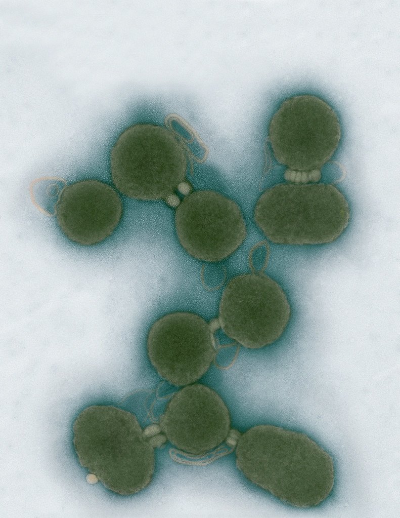
Minimal Cell — JCVI-syn3.0
Electron micrographs of clusters of JCVI-syn3.0 cells magnified about 15,000 times. This is the world’s first minimal bacterial cell. Its synthetic genome contains only 473 genes. Surprisingly, the functions of 149 of those genes are unknown. The images were made by Tom Deerinck and Mark Ellisman of the National Center for Imaging and Microscopy Research at the University of California at San Diego.
Leadership

J. Craig Venter, Ph.D.
Credit: Brett Shipe / J. Craig Venter Institute

Sanjay Vashee, Ph.D.
Credit: J. Craig Venter Institute

John Glass, Ph.D.
Credit: J. Craig Venter Institute
Scientists in the Lab

JCVI Scientists Working in Lab
Credit: J. Craig Venter Institute

JCVI Scientists Working in Lab
Credit: J. Craig Venter Institute

JCVI Scientists Working in Lab
Credit: J. Craig Venter Institute

JCVI Scientists Working in Lab
Credit: J. Craig Venter Institute

JCVI Scientists Working in Lab
Credit: J. Craig Venter Institute

JCVI Scientists Working in Lab
Credit: J. Craig Venter Institute
JCVI La Jolla Lab (Exterior)

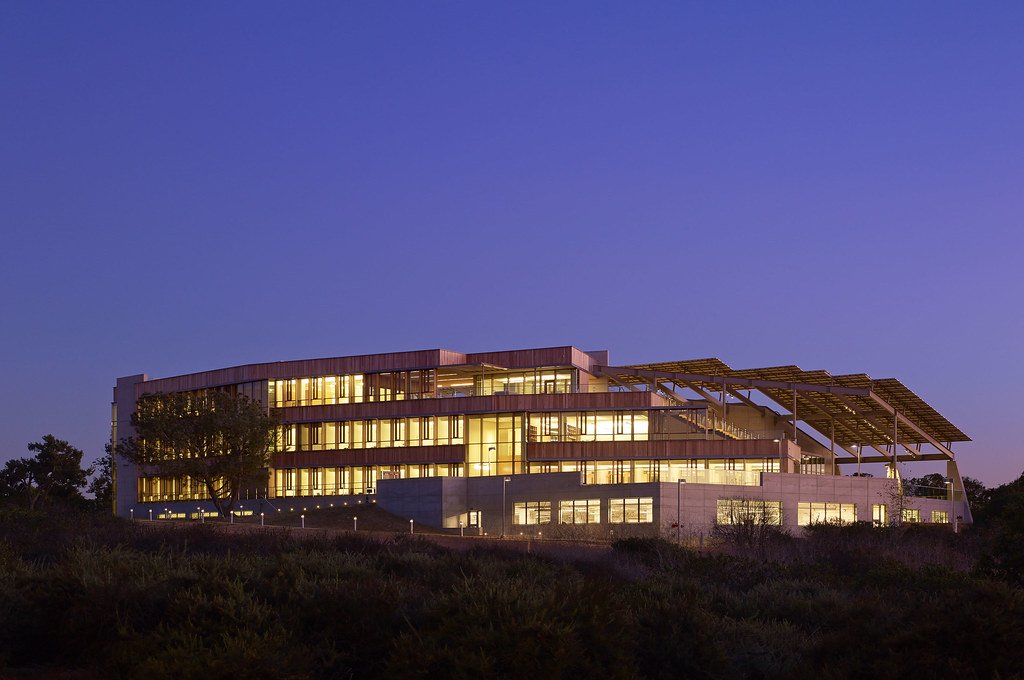
J. Craig Venter Institute, La Jolla (building exterior)
North facade at dusk. Nick Merrick © Hedrich Blessing Photographers.

J. Craig Venter Institute, La Jolla (building exterior)
South facade from soccer field. Nick Merrick © Hedrich Blessing Photographers.

J. Craig Venter Institute, La Jolla (building exterior)
Northwest view. Nick Merrick © Hedrich Blessing Photographers.

J. Craig Venter Institute, La Jolla (building exterior)
Northeast view of main entrance. Nick Merrick © Hedrich Blessing Photographers.

J. Craig Venter Institute, La Jolla (building exterior)
East facing main entrance at dusk. Nick Merrick © Hedrich Blessing Photographers.

J. Craig Venter Institute, La Jolla (building exterior)
East facing main entrance. Nick Merrick © Hedrich Blessing Photographers.

J. Craig Venter Institute, La Jolla (building exterior)
Building main entrance. Nick Merrick © Hedrich Blessing Photographers.

J. Craig Venter Institute, La Jolla (building exterior)
JCVI La Jolla north facade. Nick Merrick © Hedrich Blessing Photographers.

J. Craig Venter Institute, La Jolla (building exterior)
JCVI La Jolla north facade detail. Nick Merrick © Hedrich Blessing Photographers.

J. Craig Venter Institute, La Jolla (building exterior)
Rock garden in courtyard dusk. Nick Merrick © Hedrich Blessing Photographers.

J. Craig Venter Institute, La Jolla (building exterior)
Rock garden in courtyard. Nick Merrick © Hedrich Blessing Photographers.

J. Craig Venter Institute, La Jolla (building exterior)
Rock garden in courtyard. Nick Merrick © Hedrich Blessing Photographers.

J. Craig Venter Institute, La Jolla (building exterior)
People at courtyard tables. Nick Merrick © Hedrich Blessing Photographers.

J. Craig Venter Institute, La Jolla (building exterior)
2nd floor deck. © Tim Griffith.

J. Craig Venter Institute, La Jolla (building exterior)
Looking west at dusk. Nick Merrick © Hedrich Blessing Photographers.

J. Craig Venter Institute, La Jolla (building exterior)
First floor plaza looking south. Nick Merrick © Hedrich Blessing Photographers.

J. Craig Venter Institute, La Jolla (building exterior)
East main entrance closeup. Nick Merrick © Hedrich Blessing Photographers.

J. Craig Venter Institute, La Jolla (building exterior)
Stairs in courtyard. Nick Merrick © Hedrich Blessing Photographers.

J. Craig Venter Institute, La Jolla (building exterior)
Detail of southwest corner. Nick Merrick © Hedrich Blessing Photographers.

J. Craig Venter Institute, La Jolla (building exterior)
Sunset off 3rd floor deck. © Tim Griffith.

J. Craig Venter Institute, La Jolla (building exterior)
From northwest at dusk. Nick Merrick © Hedrich Blessing Photographers.

J. Craig Venter Institute, La Jolla (building exterior)
Photovoltaics looking west towards ocean. Nick Merrick © Hedrich Blessing Photographers.
JCVI La Jolla Lab (Interior)

J. Craig Venter Institute, La Jolla (building interior)
Wet lab with people. Nick Merrick © Hedrich Blessing Photographers.

J. Craig Venter Institute, La Jolla (building interior)
Single cell analyzer with researcher. © Tim Griffith.

J. Craig Venter Institute, La Jolla (building interior)
Mili-Q water purifier. © Tim Griffith.

J. Craig Venter Institute, La Jolla (building interior)
Lab bench work. Green plugs can be seen. © Tim Griffith.

J. Craig Venter Institute, La Jolla (building interior)
Cool room. © Tim Griffith.

J. Craig Venter Institute, La Jolla (building interior)
Confocal microscope. © Tim Griffith.

J. Craig Venter Institute, La Jolla (building interior)
Anaerobic glove box. © Tim Griffith.

J. Craig Venter Institute, La Jolla (building interior)
JCVI staff at DNA sequencer. © Tim Griffith.