Media Center
The complete genome sequence of Archeoglobus fulgidus is published.
TIGR releases partial sequence of Streptococcus pneumoniae type 4 strain
TIGR releases Chromosome 2 sequence from Plasmodium falciparum, the organism responsible for malaria
The sequences from eleven plasmids of Borrelia burgdorferi are now available by ftp.
TIGR Rice Gene Index
An award has been made to Dr. Robert Fleischmann at TIGR by the NIAID under the announcement "Innovative Drug Discovery in AIDS Opportunistic Infections" for the complete genome sequenceing of a strain of Mycobacterium avium
Our Ice Maiden project page is put on the Web site.
TIGR Mouse Gene Index
New release of M. tuberculosis data available by ftp.
Helicobacter pylori genome sequence published.
Pages
Media Contact
Related
SARS-CoV-2 Mutation Tracking
The Bacterial Viral Bioinformatic Resource Center (BV-BRC) is proud to introduce a new resource with the goal of providing live tracking of SARS-CoV-2 mutations. This real-time resource will provide regular reports focused on “Variants and Lineages of Concern” (VoCs/LoCs), and will serve as an early warning system for variants that are increasing in frequency in specific geographical locations.
JCVI Scientists and Interns Dramatically Trim Proteome Analysis Costs with New Lab-on-a-Filter Process
Through a happy accident and a keen mind, JCVI intern Rodrigo Eguez realized scientists might be able to pack their own filters rather than rely on those produced commercially at a significant cost savings. While playing around in the laboratory, he inadvertently disassembled a filter device...
Unique Antibody Pattern Discovered in COVID-19 ICU Patients May Be Key to Predicting Severe Outcomes
While news of promising COVID-19 vaccine trials is heartening, the fight to control infection rates and develop effective treatments will be an ongoing challenge for science for years to come. Gene Tan, PhD and his collaborators are working on identifying...
Synthetic Cell-Powered Lotion to Manage Type 1 Diabetes
Early last year we first talked about how researchers Yo Suzuki, PhD, and John Glass, PhD at JCVI set out to eliminate the need for type 1 diabetes (T1D) patients to receive insulin injections to manage blood glucose levels through a novel approach: developing a bacterial replacement for beta...
COVID-19 Further Complicating Flu Season
While the world is rightly focused on the ongoing COVID-19 pandemic, it’s important to know that influenza is always a significant public health burden, and the combination of the pandemic and flu season could converge to become a perfect storm of infectious diseases. Influenza causes 3 to...
Sara Josephine Baker
At the beginning of the 20th century, many people remained skeptical of both germ theory and preventative medicine, but pioneering physician Dr. Sara Josephine Baker fought to revolutionize public health and is credited with saving tens of thousands of lives. After studying chemistry and...
JCVI Researchers Help Advance Our Understanding of Ocean Microbes, Developing New Tools and Protocols Through Large-Scale Study
The oceans cover over two-thirds of the Earth’s surface and contain an abundance of life including diverse populations of marine microbes. Studying the genetics, biochemistry and metabolism of these microbes has been one of JCVI’s long standing research initiatives and is...
Online Education Resources to Help With Your New “Normal”
The COVID-19 pandemic has brought many changes to our daily lives and routines, including for many of you the role of an at-home educator for your children due to open-ended school closures. While we also miss directly connecting with students from our community, JCVI remains committed...
Coronavirus Pandemic: Putting Comprehensive Genomic Data in the Hands of Frontline Researchers Worldwide is Paramount
According to the CDC, SARS-CoV-2, the virus causing COVID-19, has now been detected in more than 150 countries/locations internationally. The World Health Organization (WHO) has declared COVID-19 a pandemic, and in the United States it has been declared it a national emergency. As...
Characterization of Bacteria from the International Space Station Drinking Water
From a microbiology perspective, the International Space Station (ISS) is interesting considering its microgravity, increased radiation, low humidity and elevated carbon dioxide levels. Because of its isolation, and unique environment, it is vital to study the microorganisms that thrive there...
Pages
Scientist renowned for study of adolescent brains named president of J. Craig Venter Institute
Anders Dale says he will move roughly $10 million in NIH funding from UCSD to JCVI.
Mirror Bacteria Research Poses Significant Risks, Dozens of Scientists Warn
Synthetic biologists make artificial cells, but one particular kind isn’t worth the risk.
Can CRISPR help stop African Swine Fever?
Gene editing could create a successful vaccine to protect against the viral disease that has killed close to 2 million pigs globally since 2021.
Getting Under the Skin
Amid an insulin crisis, one project aims to engineer microscopic insulin pumps out of a skin bacterium.
Planet Microbe
There are more organisms in the sea, a vital producer of oxygen on Earth, than planets and stars in the universe.
The Next Climate Change Calamity?: We’re Ruining the Microbiome, According to Human-Genome-Pioneer Craig Venter
In a new book (coauthored with Venter), a Vanity Fair contributor presents the oceanic evidence that human activity is altering the fabric of life on a microscopic scale.
Lessons from the Minimal Cell
“Despite reducing the sequence space of possible trajectories, we conclude that streamlining does not constrain fitness evolution and diversification of populations over time. Genome minimization may even create opportunities for evolutionary exploitation of essential genes, which are commonly observed to evolve more slowly.”
Even Synthetic Life Forms With a Tiny Genome Can Evolve
By watching “minimal” cells regain the fitness they lost, researchers are testing whether a genome can be too simple to evolve.
Privacy concerns sparked by human DNA accidentally collected in studies of other species
Two research teams warn that human genomic “bycatch” can reveal private information
Pages
Logos
The JCVI logo is presented in two formats: stacked and inline. Both are acceptable, with no preference towards either. Any use of the J. Craig Venter Institute logo or name must be cleared through the JCVI Marketing and Communications team. Please submit requests to info@jcvi.org.
To download, choose a version below, right-click, and select “save link as” or similar.
Images
Following are images of our facilities, research areas, and staff for use in news media, education, and noncommercial applications, given attribution noted with each image. If you require something that is not provided or would like to use the image in a commercial application please reach out to the JCVI Marketing and Communications team at info@jcvi.org.
Human Genome

The Diploid Genome Sequence of J. Craig Venter
gff2ps achieved another genome landmark to visualize the annotation of the first published human diploid genome, included as Poster S1 of “The Diploid Genome Sequence of J. Craig Venter” (Levy et al., PLoS Biology, 5(10):e254, 2007). Courtesy J.F. Abril / Computational Genomics Lab, Universitat de Barcelona (compgen.bio.ub.edu/Genome_Posters).

Annotation of the Celera Human Genome Assembly
We have drawn the map of the Human Genome with gff2ps. 22 autosomic, X and Y chromosomes were displayed in a big poster appearing as Figure 1 of “The Sequence of the Human Genome” (Venter et al., Science, 291(5507):1304-1351, 2001). The single chromosome pictures can be accessed from here to visualize the web version of the “Annotation of the Celera Human Genome Assembly” poster. Courtesy J.F. Abril / Computational Genomics Lab, Universitat de Barcelona (compgen.bio.ub.edu/Genome_Posters).
Synthetic Cell

J. Craig Venter, Ph.D. and Hamilton O. Smith, M.D.
Credit: J. Craig Venter Institute

Hamilton O. Smith, M.D. and Clyde A. Hutchison III, Ph.D.
Credit: J. Craig Venter Institute

J. Craig Venter, Ph.D.
Credit: Brett Shipe / J. Craig Venter Institute

Clyde A. Hutchison III, Ph.D.
Credit: J. Craig Venter Institute

John Glass, Ph.D.
Credit: J. Craig Venter Institute

Dan Gibson, Ph.D.
Credit: J. Craig Venter Institute

Carole Lartigue, Ph.D.
Credit: J. Craig Venter Institute

JCVI Synthetic Biology Team
Credit: J. Craig Venter Institute

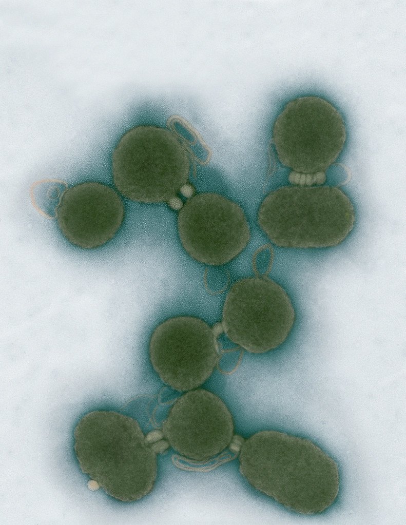
Aggregated M. mycoides JCVI-syn1.0
Negatively stained transmission electron micrographs of aggregated M. mycoides JCVI-syn1.0. Cells using 1% uranyl acetate on pure carbon substrate visualized using JEOL 1200EX transmission electron microscope at 80 keV. Electron micrographs were provided by Tom Deerinck and Mark Ellisman of the National Center for Microscopy and Imaging Research at the University of California at San Diego.

Dividing M. mycoides JCVI-syn1.0
Negatively stained transmission electron micrographs of dividing M. mycoides JCVI-syn1.0. Freshly fixed cells were stained using 1% uranyl acetate on pure carbon substrate visualized using JEOL 1200EX transmission electron microscope at 80 keV. Electron micrographs were provided by Tom Deerinck and Mark Ellisman of the National Center for Microscopy and Imaging Research at the University of California at San Diego.

Scanning Electron Micrographs of M. mycoides JCVI-syn1
Scanning electron micrographs of M. mycoides JCVI-syn1. Samples were post-fixed in osmium tetroxide, dehydrated and critical point dried with CO2 , then visualized using a Hitachi SU6600 scanning electron microscope at 2.0 keV. Electron micrographs were provided by Tom Deerinck and Mark Ellisman of the National Center for Microscopy and Imaging Research at the University of California at San Diego.

Mycoplasma mycoides JCVI-syn1.0
Credit: J. Craig Venter Institute

The Assembly of a Synthetic M. mycoides Genome in Yeast
Credit: J. Craig Venter Institute

M. mycoides JCVI-syn 1.0 and WT M. mycoides
Credit: J. Craig Venter Institute

Creating Bacteria from Prokaryotic Genomes Engineered in Yeast
Credit: J. Craig Venter Institute
See more on the first self-replicating synthetic bacterial cell.
Minimal Cell

Minimal Cell — JCVI-syn3.0
Electron micrographs of clusters of JCVI-syn3.0 cells magnified about 15,000 times. This is the world’s first minimal bacterial cell. Its synthetic genome contains only 473 genes. Surprisingly, the functions of 149 of those genes are unknown. The images were made by Tom Deerinck and Mark Ellisman of the National Center for Imaging and Microscopy Research at the University of California at San Diego.
Leadership

J. Craig Venter, Ph.D.
Credit: Brett Shipe / J. Craig Venter Institute

Sanjay Vashee, Ph.D.
Credit: J. Craig Venter Institute

John Glass, Ph.D.
Credit: J. Craig Venter Institute
Scientists in the Lab

JCVI Scientists Working in Lab
Credit: J. Craig Venter Institute

JCVI Scientists Working in Lab
Credit: J. Craig Venter Institute

JCVI Scientists Working in Lab
Credit: J. Craig Venter Institute

JCVI Scientists Working in Lab
Credit: J. Craig Venter Institute

JCVI Scientists Working in Lab
Credit: J. Craig Venter Institute

JCVI Scientists Working in Lab
Credit: J. Craig Venter Institute
JCVI La Jolla Lab (Exterior)

J. Craig Venter Institute, La Jolla (building exterior)
North facade at dusk. Nick Merrick © Hedrich Blessing Photographers.

J. Craig Venter Institute, La Jolla (building exterior)
South facade from soccer field. Nick Merrick © Hedrich Blessing Photographers.

J. Craig Venter Institute, La Jolla (building exterior)
Northwest view. Nick Merrick © Hedrich Blessing Photographers.

J. Craig Venter Institute, La Jolla (building exterior)
Northeast view of main entrance. Nick Merrick © Hedrich Blessing Photographers.

J. Craig Venter Institute, La Jolla (building exterior)
East facing main entrance at dusk. Nick Merrick © Hedrich Blessing Photographers.

J. Craig Venter Institute, La Jolla (building exterior)
East facing main entrance. Nick Merrick © Hedrich Blessing Photographers.

J. Craig Venter Institute, La Jolla (building exterior)
Building main entrance. Nick Merrick © Hedrich Blessing Photographers.

J. Craig Venter Institute, La Jolla (building exterior)
JCVI La Jolla north facade. Nick Merrick © Hedrich Blessing Photographers.

J. Craig Venter Institute, La Jolla (building exterior)
JCVI La Jolla north facade detail. Nick Merrick © Hedrich Blessing Photographers.

J. Craig Venter Institute, La Jolla (building exterior)
Rock garden in courtyard dusk. Nick Merrick © Hedrich Blessing Photographers.

J. Craig Venter Institute, La Jolla (building exterior)
Rock garden in courtyard. Nick Merrick © Hedrich Blessing Photographers.

J. Craig Venter Institute, La Jolla (building exterior)
Rock garden in courtyard. Nick Merrick © Hedrich Blessing Photographers.

J. Craig Venter Institute, La Jolla (building exterior)
People at courtyard tables. Nick Merrick © Hedrich Blessing Photographers.

J. Craig Venter Institute, La Jolla (building exterior)
2nd floor deck. © Tim Griffith.

J. Craig Venter Institute, La Jolla (building exterior)
Looking west at dusk. Nick Merrick © Hedrich Blessing Photographers.

J. Craig Venter Institute, La Jolla (building exterior)
First floor plaza looking south. Nick Merrick © Hedrich Blessing Photographers.

J. Craig Venter Institute, La Jolla (building exterior)
East main entrance closeup. Nick Merrick © Hedrich Blessing Photographers.

J. Craig Venter Institute, La Jolla (building exterior)
Stairs in courtyard. Nick Merrick © Hedrich Blessing Photographers.

J. Craig Venter Institute, La Jolla (building exterior)
Detail of southwest corner. Nick Merrick © Hedrich Blessing Photographers.

J. Craig Venter Institute, La Jolla (building exterior)
Sunset off 3rd floor deck. © Tim Griffith.

J. Craig Venter Institute, La Jolla (building exterior)
From northwest at dusk. Nick Merrick © Hedrich Blessing Photographers.

J. Craig Venter Institute, La Jolla (building exterior)
Photovoltaics looking west towards ocean. Nick Merrick © Hedrich Blessing Photographers.
JCVI La Jolla Lab (Interior)

J. Craig Venter Institute, La Jolla (building interior)
Wet lab with people. Nick Merrick © Hedrich Blessing Photographers.

J. Craig Venter Institute, La Jolla (building interior)
Single cell analyzer with researcher. © Tim Griffith.

J. Craig Venter Institute, La Jolla (building interior)
Mili-Q water purifier. © Tim Griffith.

J. Craig Venter Institute, La Jolla (building interior)
Lab bench work. Green plugs can be seen. © Tim Griffith.

J. Craig Venter Institute, La Jolla (building interior)
Cool room. © Tim Griffith.

J. Craig Venter Institute, La Jolla (building interior)
Confocal microscope. © Tim Griffith.

J. Craig Venter Institute, La Jolla (building interior)
Anaerobic glove box. © Tim Griffith.

J. Craig Venter Institute, La Jolla (building interior)
JCVI staff at DNA sequencer. © Tim Griffith.