Media Center
JCVI Associate Professor Marcelo Freire elected to the 2022 class of AAAS Fellows
More Than 500 Scientists and Engineers Bestowed Lifetime AAAS Fellows Honor
Therapeutic Potential of Bizarre ‘Jumbo’ Viruses Tapped for $10M HHMI Emerging Pathogens Project
UC San Diego leads initiative aiming to develop bacteriophages as solutions for antibiotic resistance crisis
HHMI’s Emerging Pathogens Initiative Aims for Scientific Head Start on Future Epidemics
Synthetic genomics advances and promise
Advances in DNA synthesis will enable extraordinary new opportunities in medicine, industry, agriculture, and research
Scientists announce comprehensive regional diagnostic of microbial ocean life using DNA testing
Large-scale ‘metabarcoding’ methods could revolutionize how society understands forces that drive seafood supply, planet’s ability to remove greenhouse gases
J. Craig Venter Institute sells La Jolla laboratory building to UC San Diego
2022 Fellows of the AACR Academy will be honored during Sunday’s Opening Session
JCVI Professor Emeritus Hamilton O. Smith, MD among the inductees
Scientists develop most complete whole-cell computer simulation model of cell to date
J. Craig Venter Institute model organism-minimal cell platform provides robust tools for exploring first principles of life, design tools for genome
Omicron and Beta variants evade antibodies elicited by vaccines and previous infections, but boosters help
Pregnancy also contributes to a reduced COVID-19 antibody response
Pages
Media Contact
Related
Supporting earthquake relief efforts in Turkey and Syria
We are devastated by the recent earthquakes which have caused enormous destruction in Turkey and Syria and encourage all who are able to support organizations involved in relief efforts. Locally, the American Turkish Association of Southern California (ATASC) is raising funds and...
Leg 2: exploring the Mid-Cayman Spreading Center
Editor’s note JCVI Staff Scientist Erin Garza, Ph.D., was selected to embark on a unique research expedition aboard the HOV Alvin submersible, a crewed deep-ocean research vessel owned by the United States Navy and operated by the Woods Hole Oceanographic Institution, that has brought...
The dive: searching for deep ocean plastics in the Puerto Rico Trench
Editor’s note JCVI Staff Scientist Erin Garza, Ph.D., was selected to embark on a unique research expedition aboard the HOV Alvin submersible, a crewed deep-ocean research vessel owned by the United States Navy and operated by the Woods Hole Oceanographic Institution, that has brought...
Leg 1: headed to an unexplored area of the Puerto Rico Trench
Editor’s note JCVI Staff Scientist Erin Garza, Ph.D., was selected to embark on a unique research expedition aboard the HOV Alvin submersible, a crewed deep-ocean research vessel owned by the United States Navy and operated by the Woods Hole Oceanographic Institution, that has brought...
My journey begins: heading to the Puerto Rico Trench in search of deep-sea plastic
Editor’s note JCVI Staff Scientist Erin Garza, Ph.D., was selected to embark on a unique research expedition aboard the HOV Alvin submersible, a crewed deep-ocean research vessel owned by the United States Navy and operated by the Woods Hole Oceanographic Institution, that has brought...
Celebrating pioneers in science and medicine this Black History Month
Happy Black History Month! At JCVI, we believe in the importance of celebrating scientific trailblazers, particularly those who made groundbreaking advancements all while overcoming overt racism. Here, we have highlighted the stories and achievements of some of the most accomplished Black...
Eleven female scientists whose research changed the world
Today is Women’s Equality Day and to celebrate, we are highlighting accomplishments made by women in science and technology. While these scientists were influential in advancing their fields and championing the fair treatment of women in science, currently women only make up 28% of the...
Complete Genome Sequence of Strain JB001, a Member of Saccharibacteria Clade G6
The complexity and diversity of the microbial world was not fully understood until sequencing technology allowed us to study microbes without growing them in the lab. An important family of bacteria, Saccharibacteria (formerly called TM7), is one of the many bacteria of interest which were...
Scientific Pioneers
JCVI recognizes trailblazers in scientific history, particularly those who made advancements all while surpassing gender, ethnic, and other societal barriers, creating opportunity for the next generation of scientists. These historical figures not only helped advance our understanding of human...
Women’s History Month: Tu Youyou
Tu Youyou is a Chinese pharmaceutical chemist whose unique training in the classification of medical plants and their active ingredients resulted in a discovery that has led to the survival and improved health of millions of people. In 1967, at the height of the Vietnam War, malaria spread by...
Pages
Leonardo Da Vinci: New family tree spans 21 generations, 690 years, finds 14 living male descendants
The surprising results of a decade-long investigation by Alessandro Vezzosi and Agnese Sabato provide a strong basis for advancing a project researching Leonardo da Vinci's DNA.
Genome Research Papers on Meningococcal Recombination, Psoriasis Variants in China, More
Sailing the Seas in Search of Microbes
Projects aimed at collecting big data about the ocean’s tiniest life forms continue to expand our view of the seas.
What the Public Should Not Know
J. Craig Venter, PhD, argues scientists have “a moral obligation to communicate what they're doing to the public,” and that more studies deserve greater public criticism.
Scientists coax cells with the world’s smallest genomes to reproduce normally
The discovery could sharpen scientists’ understanding of which functions are crucial for normal cells and what the many mysterious genes in these organisms are doing
San Diego arts, health, science and youth groups to share $71M from Prebys Foundation
The J. Craig Venter Institute is the recipient of three awards totaling more than $1.5M to study SARS-CoV-2 and heart disease
Reflections on the 20th Anniversary of the First Publication of the Human Genome
A new wave of research is needed to make ample use of humanity’s “most wondrous map”
Scientists rush to determine if mutant strain of coronavirus will deepen pandemic
U.S. researchers have been slow to perform the genetic sequencing that will help clarify the situation
Pages
Logos
The JCVI logo is presented in two formats: stacked and inline. Both are acceptable, with no preference towards either. Any use of the J. Craig Venter Institute logo or name must be cleared through the JCVI Marketing and Communications team. Please submit requests to info@jcvi.org.
To download, choose a version below, right-click, and select “save link as” or similar.
Images
Following are images of our facilities, research areas, and staff for use in news media, education, and noncommercial applications, given attribution noted with each image. If you require something that is not provided or would like to use the image in a commercial application please reach out to the JCVI Marketing and Communications team at info@jcvi.org.
Human Genome

The Diploid Genome Sequence of J. Craig Venter
gff2ps achieved another genome landmark to visualize the annotation of the first published human diploid genome, included as Poster S1 of “The Diploid Genome Sequence of J. Craig Venter” (Levy et al., PLoS Biology, 5(10):e254, 2007). Courtesy J.F. Abril / Computational Genomics Lab, Universitat de Barcelona (compgen.bio.ub.edu/Genome_Posters).

Annotation of the Celera Human Genome Assembly
We have drawn the map of the Human Genome with gff2ps. 22 autosomic, X and Y chromosomes were displayed in a big poster appearing as Figure 1 of “The Sequence of the Human Genome” (Venter et al., Science, 291(5507):1304-1351, 2001). The single chromosome pictures can be accessed from here to visualize the web version of the “Annotation of the Celera Human Genome Assembly” poster. Courtesy J.F. Abril / Computational Genomics Lab, Universitat de Barcelona (compgen.bio.ub.edu/Genome_Posters).
Synthetic Cell

J. Craig Venter, Ph.D. and Hamilton O. Smith, M.D.
Credit: J. Craig Venter Institute

Hamilton O. Smith, M.D. and Clyde A. Hutchison III, Ph.D.
Credit: J. Craig Venter Institute

J. Craig Venter, Ph.D.
Credit: Brett Shipe / J. Craig Venter Institute

Clyde A. Hutchison III, Ph.D.
Credit: J. Craig Venter Institute

John Glass, Ph.D.
Credit: J. Craig Venter Institute

Dan Gibson, Ph.D.
Credit: J. Craig Venter Institute

Carole Lartigue, Ph.D.
Credit: J. Craig Venter Institute

JCVI Synthetic Biology Team
Credit: J. Craig Venter Institute

Aggregated M. mycoides JCVI-syn1.0
Negatively stained transmission electron micrographs of aggregated M. mycoides JCVI-syn1.0. Cells using 1% uranyl acetate on pure carbon substrate visualized using JEOL 1200EX transmission electron microscope at 80 keV. Electron micrographs were provided by Tom Deerinck and Mark Ellisman of the National Center for Microscopy and Imaging Research at the University of California at San Diego.

Dividing M. mycoides JCVI-syn1.0
Negatively stained transmission electron micrographs of dividing M. mycoides JCVI-syn1.0. Freshly fixed cells were stained using 1% uranyl acetate on pure carbon substrate visualized using JEOL 1200EX transmission electron microscope at 80 keV. Electron micrographs were provided by Tom Deerinck and Mark Ellisman of the National Center for Microscopy and Imaging Research at the University of California at San Diego.

Scanning Electron Micrographs of M. mycoides JCVI-syn1
Scanning electron micrographs of M. mycoides JCVI-syn1. Samples were post-fixed in osmium tetroxide, dehydrated and critical point dried with CO2 , then visualized using a Hitachi SU6600 scanning electron microscope at 2.0 keV. Electron micrographs were provided by Tom Deerinck and Mark Ellisman of the National Center for Microscopy and Imaging Research at the University of California at San Diego.

Mycoplasma mycoides JCVI-syn1.0
Credit: J. Craig Venter Institute

The Assembly of a Synthetic M. mycoides Genome in Yeast
Credit: J. Craig Venter Institute

M. mycoides JCVI-syn 1.0 and WT M. mycoides
Credit: J. Craig Venter Institute

Creating Bacteria from Prokaryotic Genomes Engineered in Yeast
Credit: J. Craig Venter Institute
See more on the first self-replicating synthetic bacterial cell.
Minimal Cell

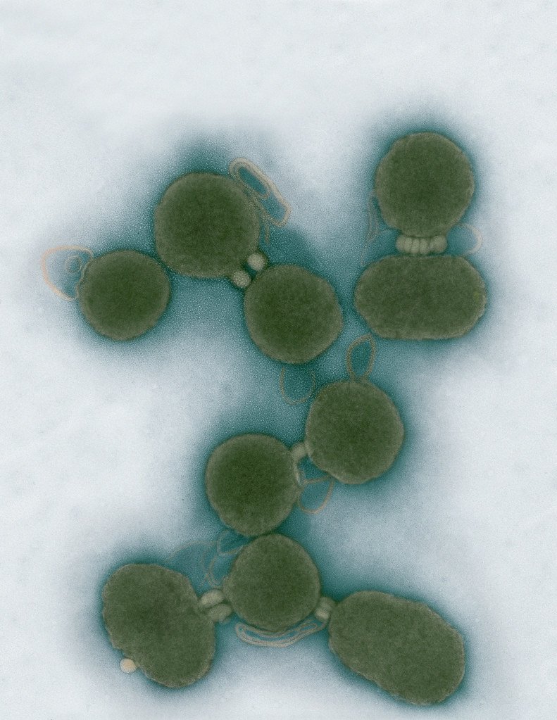
Minimal Cell — JCVI-syn3.0
Electron micrographs of clusters of JCVI-syn3.0 cells magnified about 15,000 times. This is the world’s first minimal bacterial cell. Its synthetic genome contains only 473 genes. Surprisingly, the functions of 149 of those genes are unknown. The images were made by Tom Deerinck and Mark Ellisman of the National Center for Imaging and Microscopy Research at the University of California at San Diego.
Leadership

J. Craig Venter, Ph.D.
Credit: Brett Shipe / J. Craig Venter Institute

Sanjay Vashee, Ph.D.
Credit: J. Craig Venter Institute

John Glass, Ph.D.
Credit: J. Craig Venter Institute
Scientists in the Lab

JCVI Scientists Working in Lab
Credit: J. Craig Venter Institute

JCVI Scientists Working in Lab
Credit: J. Craig Venter Institute

JCVI Scientists Working in Lab
Credit: J. Craig Venter Institute

JCVI Scientists Working in Lab
Credit: J. Craig Venter Institute

JCVI Scientists Working in Lab
Credit: J. Craig Venter Institute

JCVI Scientists Working in Lab
Credit: J. Craig Venter Institute
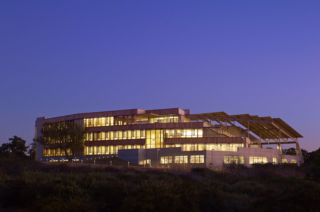
JCVI La Jolla Lab (Exterior)

J. Craig Venter Institute, La Jolla (building exterior)
North facade at dusk. Nick Merrick © Hedrich Blessing Photographers.

J. Craig Venter Institute, La Jolla (building exterior)
South facade from soccer field. Nick Merrick © Hedrich Blessing Photographers.

J. Craig Venter Institute, La Jolla (building exterior)
Northwest view. Nick Merrick © Hedrich Blessing Photographers.

J. Craig Venter Institute, La Jolla (building exterior)
Northeast view of main entrance. Nick Merrick © Hedrich Blessing Photographers.

J. Craig Venter Institute, La Jolla (building exterior)
East facing main entrance at dusk. Nick Merrick © Hedrich Blessing Photographers.

J. Craig Venter Institute, La Jolla (building exterior)
East facing main entrance. Nick Merrick © Hedrich Blessing Photographers.

J. Craig Venter Institute, La Jolla (building exterior)
Building main entrance. Nick Merrick © Hedrich Blessing Photographers.

J. Craig Venter Institute, La Jolla (building exterior)
JCVI La Jolla north facade. Nick Merrick © Hedrich Blessing Photographers.

J. Craig Venter Institute, La Jolla (building exterior)
JCVI La Jolla north facade detail. Nick Merrick © Hedrich Blessing Photographers.

J. Craig Venter Institute, La Jolla (building exterior)
Rock garden in courtyard dusk. Nick Merrick © Hedrich Blessing Photographers.

J. Craig Venter Institute, La Jolla (building exterior)
Rock garden in courtyard. Nick Merrick © Hedrich Blessing Photographers.

J. Craig Venter Institute, La Jolla (building exterior)
Rock garden in courtyard. Nick Merrick © Hedrich Blessing Photographers.

J. Craig Venter Institute, La Jolla (building exterior)
People at courtyard tables. Nick Merrick © Hedrich Blessing Photographers.

J. Craig Venter Institute, La Jolla (building exterior)
2nd floor deck. © Tim Griffith.

J. Craig Venter Institute, La Jolla (building exterior)
Looking west at dusk. Nick Merrick © Hedrich Blessing Photographers.

J. Craig Venter Institute, La Jolla (building exterior)
First floor plaza looking south. Nick Merrick © Hedrich Blessing Photographers.

J. Craig Venter Institute, La Jolla (building exterior)
East main entrance closeup. Nick Merrick © Hedrich Blessing Photographers.

J. Craig Venter Institute, La Jolla (building exterior)
Stairs in courtyard. Nick Merrick © Hedrich Blessing Photographers.

J. Craig Venter Institute, La Jolla (building exterior)
Detail of southwest corner. Nick Merrick © Hedrich Blessing Photographers.

J. Craig Venter Institute, La Jolla (building exterior)
Sunset off 3rd floor deck. © Tim Griffith.

J. Craig Venter Institute, La Jolla (building exterior)
From northwest at dusk. Nick Merrick © Hedrich Blessing Photographers.

J. Craig Venter Institute, La Jolla (building exterior)
Photovoltaics looking west towards ocean. Nick Merrick © Hedrich Blessing Photographers.
JCVI La Jolla Lab (Interior)

J. Craig Venter Institute, La Jolla (building interior)
Wet lab with people. Nick Merrick © Hedrich Blessing Photographers.

J. Craig Venter Institute, La Jolla (building interior)
Single cell analyzer with researcher. © Tim Griffith.

J. Craig Venter Institute, La Jolla (building interior)
Mili-Q water purifier. © Tim Griffith.

J. Craig Venter Institute, La Jolla (building interior)
Lab bench work. Green plugs can be seen. © Tim Griffith.

J. Craig Venter Institute, La Jolla (building interior)
Cool room. © Tim Griffith.

J. Craig Venter Institute, La Jolla (building interior)
Confocal microscope. © Tim Griffith.

J. Craig Venter Institute, La Jolla (building interior)
Anaerobic glove box. © Tim Griffith.

J. Craig Venter Institute, La Jolla (building interior)
JCVI staff at DNA sequencer. © Tim Griffith.